Visible Human Project
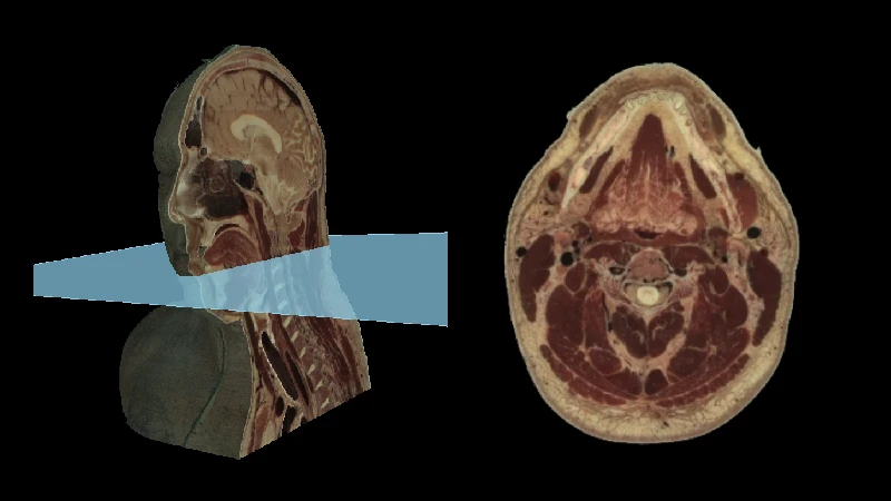
Select cases from the Visible Human Project, courtesy of the U.S. National Library of Medicine.
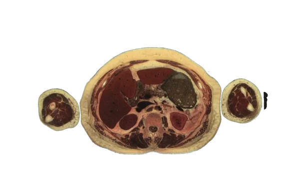
Female Chest, Abdomen and Pelvis from the Visible Human Project (Half Resolution)
Chest, abdomen and pelvis portion of the female images from the Visible Human Project, courtesy of the U.S. National Library of Medicine. The original cryo cross-section images were at 0.33 mm intervals and with each pixel 0.33 mm in size. This case has been downsampled in the z-axis by a factor of 6 and in the x- and y-axes by a factor of 2. This allows decreased file size and the ability to perform 3D processing.
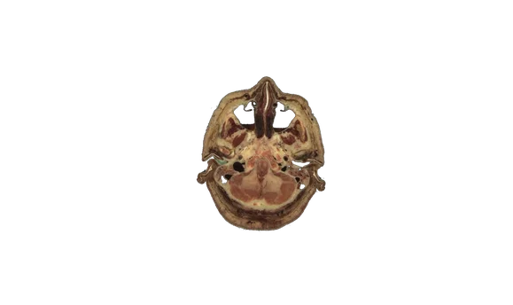
Female Head and Neck from the Visible Human Project (Half Resolution)
Head and neck portion of the female images from the Visible Human Project, courtesy of the U.S. National Library of Medicine. The original cryo cross-section images were at 0.33 mm intervals and with each pixel 0.33 mm in size. This case has been downsampled in the z-axis by a factor of 6 and in the x- and y-axes by a factor of 2. This allows decreased file size and the ability to perform 3D processing.
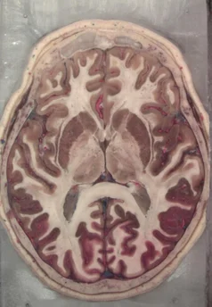
Head Anatomy Module from the Visible Human Project (Half Resolution)
Additional head images from the Visible Human Project, courtesy of the U.S. National Library of Medicine. For these images, the donor was preserved in formalin and the blood vessels were filled with araldite-F. After freezing the specimen was cryo sectioned at 0.147mm intervals and digital photographs were taken with a resolution of 1056 x 1528 pixels. This case has been downsampled by a factor of 2 from the originals for decreased file sizes and to have the ability for 3D processing. Automatic brain cortical and subcortical segmentations are performed using the Freesurfer software, in particular the SynthSeg program (for details see SynthSeg: Segmentation of brain MRI scans of any contrast and resolution without retraining. B Billot, DN Greve, O Puonti, A Thielscher, K Van Leemput, B Fischl, AV Dalca, JE Iglesias. Medical Image Analysis, 83, 102789 (2023)). The cortical segmentations were subsequently expanded for improved visibility. You can use the keyboard shortcut "a" to toggle the visibility of the segmentations.
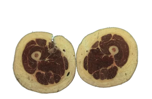
Thigh Anatomy Module from the Visible Human Project (Half Resolution)
Anatomy module made from the thigh portion of the female images from the Visible Human Project, courtesy of the U.S. National Library of Medicine. The anatomic segmentations were performed by researchers at the University of Denver and the Center for Orthopaedic Biomechanics with citation below. You may press the keyboard shortcut 'a' to toggle visibility of the color segmentations. The original cryo cross-section images were at 0.33 mm intervals and with each pixel 0.33 mm in size. This case has been downsampled in the z-axis by a factor of 6 and in the x and y-axis by a factor 2 to allow 3D processing. License: Creative Commons Attribution 4.0 International License (CC BY 4.0) for the segmentations Citations: T. E. Andreassen, D. R. Hume, L. D. Hamilton, K. E. Walker, S. E. Higinbotham, and K. B. Shelburne, “Three Dimensional Lower Extremity Musculoskeletal Geometry of the Visible Human Female and Male,” Sci. Data, vol. 10, no. 1, p. 34, Jan. 2023, doi: 10.1038/s41597-022-01905-2.
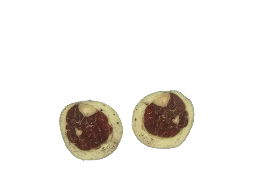
Leg Anatomy Module from the Visible Human Project (Half Resolution)
Anatomy module made from the leg portion of the female images from the Visible Human Project, courtesy of the U.S. National Library of Medicine. The anatomic segmentations were performed by researchers at the University of Denver and the Center for Orthopaedic Biomechanics with citation below. You may press the keyboard shortcut 'a' to toggle visibility of the color segmentations. The original cryo cross-section images were at 0.33 mm intervals and with each pixel 0.33 mm in size. This case has been downsampled in the z-axis by a factor of 6 and in the x and y-axis by a factor 2 to allow 3D processing. License: Creative Commons Attribution 4.0 International License (CC BY 4.0) for the segmentations Citations: T. E. Andreassen, D. R. Hume, L. D. Hamilton, K. E. Walker, S. E. Higinbotham, and K. B. Shelburne, “Three Dimensional Lower Extremity Musculoskeletal Geometry of the Visible Human Female and Male,” Sci. Data, vol. 10, no. 1, p. 34, Jan. 2023, doi: 10.1038/s41597-022-01905-2.
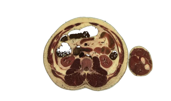
Male Chest, Abdomen and Pelvis from the Visible Human Project (Half Resolution)
Chest, abdomen and pelvis portion of the male images from the Visible Human Project, courtesy of the U.S. National Library of Medicine. The original cryo cross-section images were at 1 mm intervals and with each pixel 0.33 mm in size. This case has been downsampled by a factor of 2 from the originals for decreased file sizes and to have the ability for 3D processing.
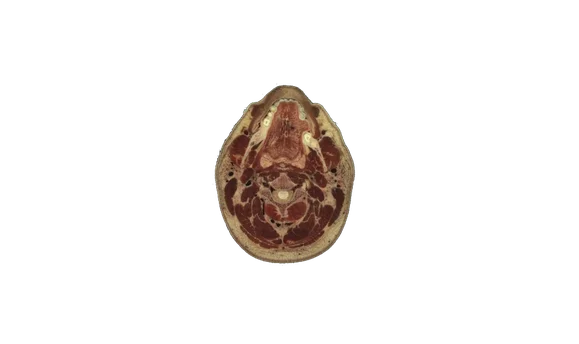
Male Head and Neck from the Visible Human Project (Half Resolution)
Head and neck portion of the male images from the Visible Human Project, courtesy of the U.S. National Library of Medicine. The original cryo cross-section images were at 1 mm intervals and with each pixel 0.33 mm in size. This case has been downsampled by a factor of 2 from the originals for decreased file sizes and to have the ability for 3D processing.
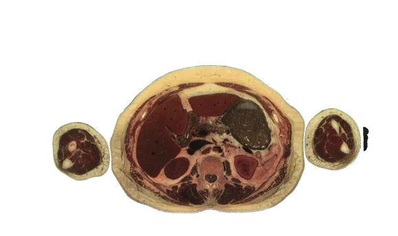
Female Chest, Abdomen and Pelvis from the Visible Human Project
Chest, abdomen and pelvis portion of the female images from the Visible Human Project, courtesy of the U.S. National Library of Medicine. The original cryo cross-section images were at 0.33 mm intervals and with each pixel 0.33 mm in size. In order to be able to fit the dataset in memory, this case has been downsampled in the z-axis by a factor of 3. The cryo sections are therefore at 1 mm intervals, similar to the male dataset. Note: Due to the large file size, attempting to perform either 3D MPR or 3D VRT on this dataset will crash your current tab's WebGL session. This would then require a full page refresh to be functional again. If you need to do 3D processing, use the "half resoltion" variant instead.
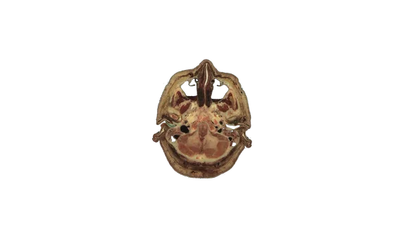
Female Head and Neck from the Visible Human Project
Head and neck portion of the female images from the Visible Human Project, courtesy of the U.S. National Library of Medicine. The original cryo cross-section images were at 0.33 mm intervals and with each pixel 0.33 mm in size. In order to be able to fit the dataset in memory, this case has been downsampled in the z-axis by a factor of 3. The cryo sections are therefore at 1 mm intervals, similar to the male dataset. Note: Due to the large file size, attempting to perform either 3D MPR or 3D VRT on this dataset will crash your current tab's WebGL session. This would then require a full page refresh to be functional again. If you need to do 3D processing, use the "half resoltion" variant instead.
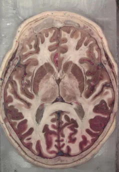
Head Anatomy Module from the Visible Human Project
Additional head images from the Visible Human Project, courtesy of the U.S. National Library of Medicine. For these images, the donor was preserved in formalin and the blood vessels were filled with araldite-F. After freezing the specimen was cryo sectioned at 0.147mm intervals and digital photographs were taken with a resolution of 1056 x 1528 pixels. Automatic brain cortical and subcortical segmentations are performed using the Freesurfer software, in particular the SynthSeg program (for details see SynthSeg: Segmentation of brain MRI scans of any contrast and resolution without retraining. B Billot, DN Greve, O Puonti, A Thielscher, K Van Leemput, B Fischl, AV Dalca, JE Iglesias. Medical Image Analysis, 83, 102789 (2023)). The cortical segmentations were subsequently expanded for improved visibility. You can use the keyboard shortcut "a" to toggle the visibility of the segmentations. Note: Due to the large file size, attempting to perform either 3D MPR or 3D VRT on this dataset will crash your current tab's WebGL session. This would then require a full page refresh to be functional again. If you need to do 3D processing, use the "half resoltion" variant instead.
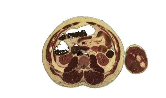
Male Chest, Abdomen and Pelvis from the Visible Human Project
Chest, abdomen and pelvis portion of the male images from the Visible Human Project, courtesy of the U.S. National Library of Medicine. The original cryo cross-section images were at 1 mm intervals and with each pixel 0.33 mm in size. Note: Due to the large file size, attempting to perform either 3D MPR or 3D VRT on this dataset will crash your current tab's WebGL session. This would then require a full page refresh to be functional again. If you need to do 3D processing, use the "half resoltion" variant instead.
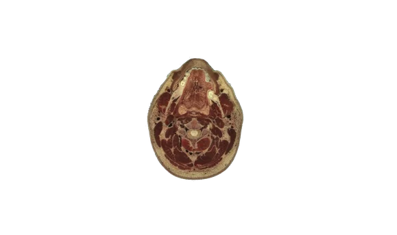
Male Head and Neck from the Visible Human Project
Head and neck portion of the male images from the Visible Human Project, courtesy of the U.S. National Library of Medicine. The original cryo cross-section images were at 1 mm intervals and with each pixel 0.33 mm in size. Note: Due to the large file size, attempting to perform either 3D MPR or 3D VRT on this dataset will crash your current tab's WebGL session. This would then require a full page refresh to be functional again. If you need to do 3D processing, use the "half resoltion" variant instead.
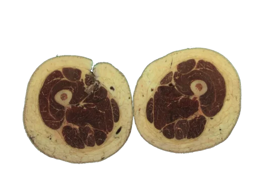
Thigh Anatomy Module from the Visible Human Project
Anatomy module made from the thigh portion of the female images from the Visible Human Project, courtesy of the U.S. National Library of Medicine. The anatomic segmentations were performed by researchers at the University of Denver and the Center for Orthopaedic Biomechanics with citation below. You may press the keyboard shortcut 'a' to toggle visibility of the color segmentations. The original cryo cross-section images were at 0.33 mm intervals and with each pixel 0.33 mm in size. In order to be able to fit the dataset in memory, this case has been downsampled in the z-axis by a factor of 3. The cryo sections are therefore at 1 mm intervals, similar to the male dataset. Note: Due to the large file size, attempting to perform either 3D MPR or 3D VRT on this dataset will crash your current tab's WebGL session. This would then require a full page refresh to be functional again. If you need to do 3D processing, use the "half resoltion" variant instead. License: Creative Commons Attribution 4.0 International License (CC BY 4.0) for the segmentations Citations: T. E. Andreassen, D. R. Hume, L. D. Hamilton, K. E. Walker, S. E. Higinbotham, and K. B. Shelburne, “Three Dimensional Lower Extremity Musculoskeletal Geometry of the Visible Human Female and Male,” Sci. Data, vol. 10, no. 1, p. 34, Jan. 2023, doi: 10.1038/s41597-022-01905-2.
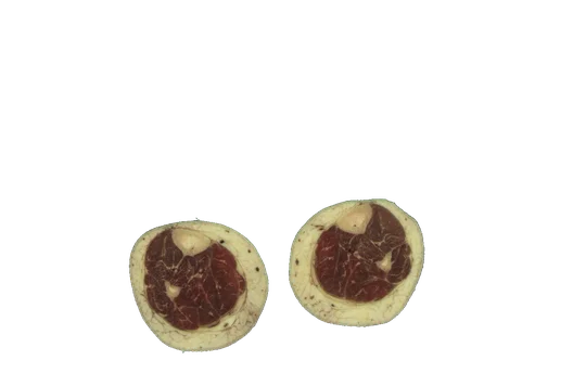
Leg Anatomy Module from the Visible Human Project
Anatomy module made from the leg portion of the female images from the Visible Human Project, courtesy of the U.S. National Library of Medicine. The anatomic segmentations were performed by researchers at the University of Denver and the Center for Orthopaedic Biomechanics with citation below. You may press the keyboard shortcut 'a' to toggle visibility of the color segmentations. The original cryo cross-section images were at 0.33 mm intervals and with each pixel 0.33 mm in size. In order to be able to fit the dataset in memory, this case has been downsampled in the z-axis by a factor of 3. The cryo sections are therefore at 1 mm intervals, similar to the male dataset. Note: Due to the large file size, attempting to perform either 3D MPR or 3D VRT on this dataset will crash your current tab's WebGL session. This would then require a full page refresh to be functional again. If you need to do 3D processing, use the "half resoltion" variant instead. License: Creative Commons Attribution 4.0 International License (CC BY 4.0) for the segmentations Citations: T. E. Andreassen, D. R. Hume, L. D. Hamilton, K. E. Walker, S. E. Higinbotham, and K. B. Shelburne, “Three Dimensional Lower Extremity Musculoskeletal Geometry of the Visible Human Female and Male,” Sci. Data, vol. 10, no. 1, p. 34, Jan. 2023, doi: 10.1038/s41597-022-01905-2.