PROSTATE-MRI: Prostate Cancer
These cases are from the PROSTATE-MRI dataset, a collection of 26 pathology proven prostate cancer cases. Each case has high resolution prostate MRI as well as radical prostatectomy histopathology images forming a valuable radiology-pathology correlative dataset. The data was generated at the National Cancer Institute, Bethesda, Maryland, USA between 2008-2010.
License: CC BY 3.0
Citation:
Choyke P, Turkbey B, Pinto P, Merino M, Wood B. (2016). Data From PROSTATE-MRI. The Cancer Imaging Archive. http://doi.org/10.7937/K9/TCIA.2016.6046GUDv
If you use this case or dataset, you must also abide by the TCIA Data Usage Policies and Restrictions.
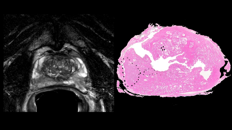
Prostate MRI - Case 0001
This is a confirmed case of prostate cancer from the PROSTATE-MRI dataset. This collection of prostate MRIs was obtained with an endorectal and phased array surface coil at 3T (Philips Achieva). Each patient had biopsy confirmation of cancer and underwent a robotic-assisted radical prostatectomy. A mold was generated from each MRI, and the prostatectomy specimen was first placed in the mold, then cut in the same plane as the MRI. The data was generated at the National Cancer Institute, Bethesda, Maryland, USA between 2008-2010.Note: The prostatectomy color images are very high resolution. Zoom in to see fine detail if interested.License: CC BY 3.0Citation: Choyke P, Turkbey B, Pinto P, Merino M, Wood B. (2016). Data From PROSTATE-MRI. The Cancer Imaging Archive. http://doi.org/10.7937/K9/TCIA.2016.6046GUDv
Prostate MRI - Case 0002
This is a confirmed case of prostate cancer from the PROSTATE-MRI dataset. This collection of prostate MRIs was obtained with an endorectal and phased array surface coil at 3T (Philips Achieva). Each patient had biopsy confirmation of cancer and underwent a robotic-assisted radical prostatectomy. A mold was generated from each MRI, and the prostatectomy specimen was first placed in the mold, then cut in the same plane as the MRI. The data was generated at the National Cancer Institute, Bethesda, Maryland, USA between 2008-2010.Note: The prostatectomy color images are very high resolution. Zoom in to see fine detail if interested.License: CC BY 3.0Citation: Choyke P, Turkbey B, Pinto P, Merino M, Wood B. (2016). Data From PROSTATE-MRI. The Cancer Imaging Archive. http://doi.org/10.7937/K9/TCIA.2016.6046GUDv
Prostate MRI - Case 0003
This is a confirmed case of prostate cancer from the PROSTATE-MRI dataset. This collection of prostate MRIs was obtained with an endorectal and phased array surface coil at 3T (Philips Achieva). Each patient had biopsy confirmation of cancer and underwent a robotic-assisted radical prostatectomy. A mold was generated from each MRI, and the prostatectomy specimen was first placed in the mold, then cut in the same plane as the MRI. The data was generated at the National Cancer Institute, Bethesda, Maryland, USA between 2008-2010.Note: The prostatectomy color images are very high resolution. Zoom in to see fine detail if interested.License: CC BY 3.0Citation: Choyke P, Turkbey B, Pinto P, Merino M, Wood B. (2016). Data From PROSTATE-MRI. The Cancer Imaging Archive. http://doi.org/10.7937/K9/TCIA.2016.6046GUDv
Prostate MRI - Case 0004
This is a confirmed case of prostate cancer from the PROSTATE-MRI dataset. This collection of prostate MRIs was obtained with an endorectal and phased array surface coil at 3T (Philips Achieva). Each patient had biopsy confirmation of cancer and underwent a robotic-assisted radical prostatectomy. A mold was generated from each MRI, and the prostatectomy specimen was first placed in the mold, then cut in the same plane as the MRI. The data was generated at the National Cancer Institute, Bethesda, Maryland, USA between 2008-2010.Note: The prostatectomy color images are very high resolution. Zoom in to see fine detail if interested.License: CC BY 3.0Citation: Choyke P, Turkbey B, Pinto P, Merino M, Wood B. (2016). Data From PROSTATE-MRI. The Cancer Imaging Archive. http://doi.org/10.7937/K9/TCIA.2016.6046GUDv
Prostate MRI - Case 0005
This is a confirmed case of prostate cancer from the PROSTATE-MRI dataset. This collection of prostate MRIs was obtained with an endorectal and phased array surface coil at 3T (Philips Achieva). Each patient had biopsy confirmation of cancer and underwent a robotic-assisted radical prostatectomy. A mold was generated from each MRI, and the prostatectomy specimen was first placed in the mold, then cut in the same plane as the MRI. The data was generated at the National Cancer Institute, Bethesda, Maryland, USA between 2008-2010.Note: The prostatectomy color images are very high resolution. Zoom in to see fine detail if interested.License: CC BY 3.0Citation: Choyke P, Turkbey B, Pinto P, Merino M, Wood B. (2016). Data From PROSTATE-MRI. The Cancer Imaging Archive. http://doi.org/10.7937/K9/TCIA.2016.6046GUDv
Prostate MRI - Case 0006
This is a confirmed case of prostate cancer from the PROSTATE-MRI dataset. This collection of prostate MRIs was obtained with an endorectal and phased array surface coil at 3T (Philips Achieva). Each patient had biopsy confirmation of cancer and underwent a robotic-assisted radical prostatectomy. A mold was generated from each MRI, and the prostatectomy specimen was first placed in the mold, then cut in the same plane as the MRI. The data was generated at the National Cancer Institute, Bethesda, Maryland, USA between 2008-2010.Note: The prostatectomy color images are very high resolution. Zoom in to see fine detail if interested.License: CC BY 3.0Citation: Choyke P, Turkbey B, Pinto P, Merino M, Wood B. (2016). Data From PROSTATE-MRI. The Cancer Imaging Archive. http://doi.org/10.7937/K9/TCIA.2016.6046GUDv
Prostate MRI - Case 0007
This is a confirmed case of prostate cancer from the PROSTATE-MRI dataset. This collection of prostate MRIs was obtained with an endorectal and phased array surface coil at 3T (Philips Achieva). Each patient had biopsy confirmation of cancer and underwent a robotic-assisted radical prostatectomy. A mold was generated from each MRI, and the prostatectomy specimen was first placed in the mold, then cut in the same plane as the MRI. The data was generated at the National Cancer Institute, Bethesda, Maryland, USA between 2008-2010.Note: The prostatectomy color images are very high resolution. Zoom in to see fine detail if interested.License: CC BY 3.0Citation: Choyke P, Turkbey B, Pinto P, Merino M, Wood B. (2016). Data From PROSTATE-MRI. The Cancer Imaging Archive. http://doi.org/10.7937/K9/TCIA.2016.6046GUDv
Prostate MRI - Case 0008
This is a confirmed case of prostate cancer from the PROSTATE-MRI dataset. This collection of prostate MRIs was obtained with an endorectal and phased array surface coil at 3T (Philips Achieva). Each patient had biopsy confirmation of cancer and underwent a robotic-assisted radical prostatectomy. A mold was generated from each MRI, and the prostatectomy specimen was first placed in the mold, then cut in the same plane as the MRI. The data was generated at the National Cancer Institute, Bethesda, Maryland, USA between 2008-2010.Note: The prostatectomy color images are very high resolution. Zoom in to see fine detail if interested.License: CC BY 3.0Citation: Choyke P, Turkbey B, Pinto P, Merino M, Wood B. (2016). Data From PROSTATE-MRI. The Cancer Imaging Archive. http://doi.org/10.7937/K9/TCIA.2016.6046GUDv
Prostate MRI - Case 0009
This is a confirmed case of prostate cancer from the PROSTATE-MRI dataset. This collection of prostate MRIs was obtained with an endorectal and phased array surface coil at 3T (Philips Achieva). Each patient had biopsy confirmation of cancer and underwent a robotic-assisted radical prostatectomy. A mold was generated from each MRI, and the prostatectomy specimen was first placed in the mold, then cut in the same plane as the MRI. The data was generated at the National Cancer Institute, Bethesda, Maryland, USA between 2008-2010.Note: The prostatectomy color images are very high resolution. Zoom in to see fine detail if interested.License: CC BY 3.0Citation: Choyke P, Turkbey B, Pinto P, Merino M, Wood B. (2016). Data From PROSTATE-MRI. The Cancer Imaging Archive. http://doi.org/10.7937/K9/TCIA.2016.6046GUDv
Prostate MRI - Case 0010
This is a confirmed case of prostate cancer from the PROSTATE-MRI dataset. This collection of prostate MRIs was obtained with an endorectal and phased array surface coil at 3T (Philips Achieva). Each patient had biopsy confirmation of cancer and underwent a robotic-assisted radical prostatectomy. A mold was generated from each MRI, and the prostatectomy specimen was first placed in the mold, then cut in the same plane as the MRI. The data was generated at the National Cancer Institute, Bethesda, Maryland, USA between 2008-2010.Note: The prostatectomy color images are very high resolution. Zoom in to see fine detail if interested.License: CC BY 3.0Citation: Choyke P, Turkbey B, Pinto P, Merino M, Wood B. (2016). Data From PROSTATE-MRI. The Cancer Imaging Archive. http://doi.org/10.7937/K9/TCIA.2016.6046GUDv
Prostate MRI - Case 0011
This is a confirmed case of prostate cancer from the PROSTATE-MRI dataset. This collection of prostate MRIs was obtained with an endorectal and phased array surface coil at 3T (Philips Achieva). Each patient had biopsy confirmation of cancer and underwent a robotic-assisted radical prostatectomy. A mold was generated from each MRI, and the prostatectomy specimen was first placed in the mold, then cut in the same plane as the MRI. The data was generated at the National Cancer Institute, Bethesda, Maryland, USA between 2008-2010.Note: The prostatectomy color images are very high resolution. Zoom in to see fine detail if interested.License: CC BY 3.0Citation: Choyke P, Turkbey B, Pinto P, Merino M, Wood B. (2016). Data From PROSTATE-MRI. The Cancer Imaging Archive. http://doi.org/10.7937/K9/TCIA.2016.6046GUDv
Prostate MRI - Case 0012
This is a confirmed case of prostate cancer from the PROSTATE-MRI dataset. This collection of prostate MRIs was obtained with an endorectal and phased array surface coil at 3T (Philips Achieva). Each patient had biopsy confirmation of cancer and underwent a robotic-assisted radical prostatectomy. A mold was generated from each MRI, and the prostatectomy specimen was first placed in the mold, then cut in the same plane as the MRI. The data was generated at the National Cancer Institute, Bethesda, Maryland, USA between 2008-2010.Note: The prostatectomy color images are very high resolution. Zoom in to see fine detail if interested.License: CC BY 3.0Citation: Choyke P, Turkbey B, Pinto P, Merino M, Wood B. (2016). Data From PROSTATE-MRI. The Cancer Imaging Archive. http://doi.org/10.7937/K9/TCIA.2016.6046GUDv
Prostate MRI - Case 0013
This is a confirmed case of prostate cancer from the PROSTATE-MRI dataset. This collection of prostate MRIs was obtained with an endorectal and phased array surface coil at 3T (Philips Achieva). Each patient had biopsy confirmation of cancer and underwent a robotic-assisted radical prostatectomy. A mold was generated from each MRI, and the prostatectomy specimen was first placed in the mold, then cut in the same plane as the MRI. The data was generated at the National Cancer Institute, Bethesda, Maryland, USA between 2008-2010.Note: The prostatectomy color images are very high resolution. Zoom in to see fine detail if interested.License: CC BY 3.0Citation: Choyke P, Turkbey B, Pinto P, Merino M, Wood B. (2016). Data From PROSTATE-MRI. The Cancer Imaging Archive. http://doi.org/10.7937/K9/TCIA.2016.6046GUDv
Prostate MRI - Case 0014
This is a confirmed case of prostate cancer from the PROSTATE-MRI dataset. This collection of prostate MRIs was obtained with an endorectal and phased array surface coil at 3T (Philips Achieva). Each patient had biopsy confirmation of cancer and underwent a robotic-assisted radical prostatectomy. A mold was generated from each MRI, and the prostatectomy specimen was first placed in the mold, then cut in the same plane as the MRI. The data was generated at the National Cancer Institute, Bethesda, Maryland, USA between 2008-2010.Note: The prostatectomy color images are very high resolution. Zoom in to see fine detail if interested.License: CC BY 3.0Citation: Choyke P, Turkbey B, Pinto P, Merino M, Wood B. (2016). Data From PROSTATE-MRI. The Cancer Imaging Archive. http://doi.org/10.7937/K9/TCIA.2016.6046GUDv
Prostate MRI - Case 0015
This is a confirmed case of prostate cancer from the PROSTATE-MRI dataset. This collection of prostate MRIs was obtained with an endorectal and phased array surface coil at 3T (Philips Achieva). Each patient had biopsy confirmation of cancer and underwent a robotic-assisted radical prostatectomy. A mold was generated from each MRI, and the prostatectomy specimen was first placed in the mold, then cut in the same plane as the MRI. The data was generated at the National Cancer Institute, Bethesda, Maryland, USA between 2008-2010.Note: The prostatectomy color images are very high resolution. Zoom in to see fine detail if interested.License: CC BY 3.0Citation: Choyke P, Turkbey B, Pinto P, Merino M, Wood B. (2016). Data From PROSTATE-MRI. The Cancer Imaging Archive. http://doi.org/10.7937/K9/TCIA.2016.6046GUDv
Prostate MRI - Case 0016
This is a confirmed case of prostate cancer from the PROSTATE-MRI dataset. This collection of prostate MRIs was obtained with an endorectal and phased array surface coil at 3T (Philips Achieva). Each patient had biopsy confirmation of cancer and underwent a robotic-assisted radical prostatectomy. A mold was generated from each MRI, and the prostatectomy specimen was first placed in the mold, then cut in the same plane as the MRI. The data was generated at the National Cancer Institute, Bethesda, Maryland, USA between 2008-2010.Note: The prostatectomy color images are very high resolution. Zoom in to see fine detail if interested.License: CC BY 3.0Citation: Choyke P, Turkbey B, Pinto P, Merino M, Wood B. (2016). Data From PROSTATE-MRI. The Cancer Imaging Archive. http://doi.org/10.7937/K9/TCIA.2016.6046GUDv
Prostate MRI - Case 0017
This is a confirmed case of prostate cancer from the PROSTATE-MRI dataset. This collection of prostate MRIs was obtained with an endorectal and phased array surface coil at 3T (Philips Achieva). Each patient had biopsy confirmation of cancer and underwent a robotic-assisted radical prostatectomy. A mold was generated from each MRI, and the prostatectomy specimen was first placed in the mold, then cut in the same plane as the MRI. The data was generated at the National Cancer Institute, Bethesda, Maryland, USA between 2008-2010.Note: The prostatectomy color images are very high resolution. Zoom in to see fine detail if interested.License: CC BY 3.0Citation: Choyke P, Turkbey B, Pinto P, Merino M, Wood B. (2016). Data From PROSTATE-MRI. The Cancer Imaging Archive. http://doi.org/10.7937/K9/TCIA.2016.6046GUDv
Prostate MRI - Case 0018
This is a confirmed case of prostate cancer from the PROSTATE-MRI dataset. This collection of prostate MRIs was obtained with an endorectal and phased array surface coil at 3T (Philips Achieva). Each patient had biopsy confirmation of cancer and underwent a robotic-assisted radical prostatectomy. A mold was generated from each MRI, and the prostatectomy specimen was first placed in the mold, then cut in the same plane as the MRI. The data was generated at the National Cancer Institute, Bethesda, Maryland, USA between 2008-2010.Note: The prostatectomy color images are very high resolution. Zoom in to see fine detail if interested.License: CC BY 3.0Citation: Choyke P, Turkbey B, Pinto P, Merino M, Wood B. (2016). Data From PROSTATE-MRI. The Cancer Imaging Archive. http://doi.org/10.7937/K9/TCIA.2016.6046GUDv
Prostate MRI - Case 0019
This is a confirmed case of prostate cancer from the PROSTATE-MRI dataset. This collection of prostate MRIs was obtained with an endorectal and phased array surface coil at 3T (Philips Achieva). Each patient had biopsy confirmation of cancer and underwent a robotic-assisted radical prostatectomy. A mold was generated from each MRI, and the prostatectomy specimen was first placed in the mold, then cut in the same plane as the MRI. The data was generated at the National Cancer Institute, Bethesda, Maryland, USA between 2008-2010.Note: The prostatectomy color images are very high resolution. Zoom in to see fine detail if interested.License: CC BY 3.0Citation: Choyke P, Turkbey B, Pinto P, Merino M, Wood B. (2016). Data From PROSTATE-MRI. The Cancer Imaging Archive. http://doi.org/10.7937/K9/TCIA.2016.6046GUDv
Prostate MRI - Case 0020
This is a confirmed case of prostate cancer from the PROSTATE-MRI dataset. This collection of prostate MRIs was obtained with an endorectal and phased array surface coil at 3T (Philips Achieva). Each patient had biopsy confirmation of cancer and underwent a robotic-assisted radical prostatectomy. A mold was generated from each MRI, and the prostatectomy specimen was first placed in the mold, then cut in the same plane as the MRI. The data was generated at the National Cancer Institute, Bethesda, Maryland, USA between 2008-2010.Note: The prostatectomy color images are very high resolution. Zoom in to see fine detail if interested.License: CC BY 3.0Citation: Choyke P, Turkbey B, Pinto P, Merino M, Wood B. (2016). Data From PROSTATE-MRI. The Cancer Imaging Archive. http://doi.org/10.7937/K9/TCIA.2016.6046GUDv
Prostate MRI - Case 0021
This is a confirmed case of prostate cancer from the PROSTATE-MRI dataset. This collection of prostate MRIs was obtained with an endorectal and phased array surface coil at 3T (Philips Achieva). Each patient had biopsy confirmation of cancer and underwent a robotic-assisted radical prostatectomy. A mold was generated from each MRI, and the prostatectomy specimen was first placed in the mold, then cut in the same plane as the MRI. The data was generated at the National Cancer Institute, Bethesda, Maryland, USA between 2008-2010.Note: The prostatectomy color images are very high resolution. Zoom in to see fine detail if interested.License: CC BY 3.0Citation: Choyke P, Turkbey B, Pinto P, Merino M, Wood B. (2016). Data From PROSTATE-MRI. The Cancer Imaging Archive. http://doi.org/10.7937/K9/TCIA.2016.6046GUDv
Prostate MRI - Case 0022
This is a confirmed case of prostate cancer from the PROSTATE-MRI dataset. This collection of prostate MRIs was obtained with an endorectal and phased array surface coil at 3T (Philips Achieva). Each patient had biopsy confirmation of cancer and underwent a robotic-assisted radical prostatectomy. A mold was generated from each MRI, and the prostatectomy specimen was first placed in the mold, then cut in the same plane as the MRI. The data was generated at the National Cancer Institute, Bethesda, Maryland, USA between 2008-2010.Note: The prostatectomy color images are very high resolution. Zoom in to see fine detail if interested.License: CC BY 3.0Citation: Choyke P, Turkbey B, Pinto P, Merino M, Wood B. (2016). Data From PROSTATE-MRI. The Cancer Imaging Archive. http://doi.org/10.7937/K9/TCIA.2016.6046GUDv
Prostate MRI - Case 0023
This is a confirmed case of prostate cancer from the PROSTATE-MRI dataset. This collection of prostate MRIs was obtained with an endorectal and phased array surface coil at 3T (Philips Achieva). Each patient had biopsy confirmation of cancer and underwent a robotic-assisted radical prostatectomy. A mold was generated from each MRI, and the prostatectomy specimen was first placed in the mold, then cut in the same plane as the MRI. The data was generated at the National Cancer Institute, Bethesda, Maryland, USA between 2008-2010.Note: The prostatectomy color images are very high resolution. Zoom in to see fine detail if interested.License: CC BY 3.0Citation: Choyke P, Turkbey B, Pinto P, Merino M, Wood B. (2016). Data From PROSTATE-MRI. The Cancer Imaging Archive. http://doi.org/10.7937/K9/TCIA.2016.6046GUDv
Prostate MRI - Case 0024
This is a confirmed case of prostate cancer from the PROSTATE-MRI dataset. This collection of prostate MRIs was obtained with an endorectal and phased array surface coil at 3T (Philips Achieva). Each patient had biopsy confirmation of cancer and underwent a robotic-assisted radical prostatectomy. A mold was generated from each MRI, and the prostatectomy specimen was first placed in the mold, then cut in the same plane as the MRI. The data was generated at the National Cancer Institute, Bethesda, Maryland, USA between 2008-2010.Note: The prostatectomy color images are very high resolution. Zoom in to see fine detail if interested.License: CC BY 3.0Citation: Choyke P, Turkbey B, Pinto P, Merino M, Wood B. (2016). Data From PROSTATE-MRI. The Cancer Imaging Archive. http://doi.org/10.7937/K9/TCIA.2016.6046GUDv
Prostate MRI - Case 0025
This is a confirmed case of prostate cancer from the PROSTATE-MRI dataset. This collection of prostate MRIs was obtained with an endorectal and phased array surface coil at 3T (Philips Achieva). Each patient had biopsy confirmation of cancer and underwent a robotic-assisted radical prostatectomy. A mold was generated from each MRI, and the prostatectomy specimen was first placed in the mold, then cut in the same plane as the MRI. The data was generated at the National Cancer Institute, Bethesda, Maryland, USA between 2008-2010.Note: The prostatectomy color images are very high resolution. Zoom in to see fine detail if interested.License: CC BY 3.0Citation: Choyke P, Turkbey B, Pinto P, Merino M, Wood B. (2016). Data From PROSTATE-MRI. The Cancer Imaging Archive. http://doi.org/10.7937/K9/TCIA.2016.6046GUDv
Prostate MRI - Case 0026
This is a confirmed case of prostate cancer from the PROSTATE-MRI dataset. This collection of prostate MRIs was obtained with an endorectal and phased array surface coil at 3T (Philips Achieva). Each patient had biopsy confirmation of cancer and underwent a robotic-assisted radical prostatectomy. A mold was generated from each MRI, and the prostatectomy specimen was first placed in the mold, then cut in the same plane as the MRI. The data was generated at the National Cancer Institute, Bethesda, Maryland, USA between 2008-2010.Note: The prostatectomy color images are very high resolution. Zoom in to see fine detail if interested.License: CC BY 3.0Citation: Choyke P, Turkbey B, Pinto P, Merino M, Wood B. (2016). Data From PROSTATE-MRI. The Cancer Imaging Archive. http://doi.org/10.7937/K9/TCIA.2016.6046GUDv