PETWB-REP: PET/CTs with full radiology reports
Cases from PETWB-REP, a curated dataset of whole-body 18F-FDG PET/CT scans and corresponding radiology reports from 490 patients with a broad spectrum of malignancies. The data were retrospectively collected from patients who underwent clinically indicated whole-body 18F-FDG PET/CT scans at the Shanghai Universal Medical Imaging Diagnostic Center between 2021 and 2024.
License: Creative Commons Attribution 4.0 International (CC BY 4.0)
Citation:
Xue, L., Feng, G., Wenbo, Z., Zhang, Y., Li, L., Wang, S., Peng, L., Peng, S., & Gao, X. (2026). PETWB-REP: A Multi-Cancer Whole-Body FDG PET/CT Dataset with Corresponding Radiology Reports [Data set]. Zenodo. https://doi.org/10.5281/zenodo.18670487
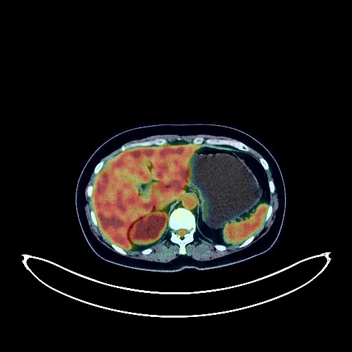
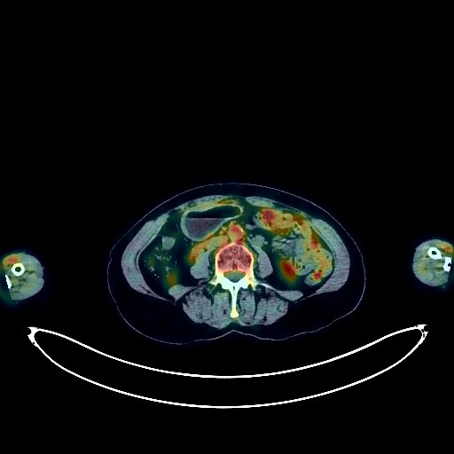
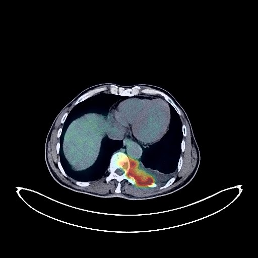
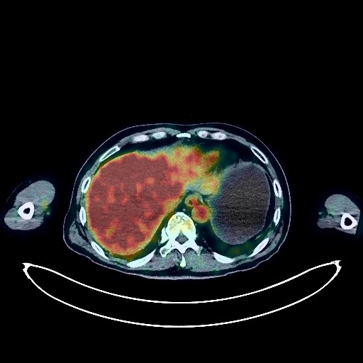
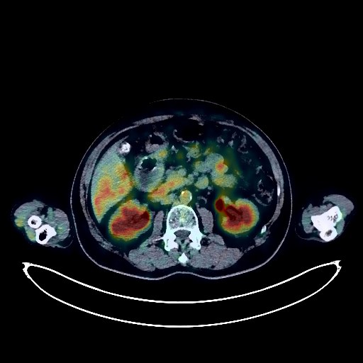
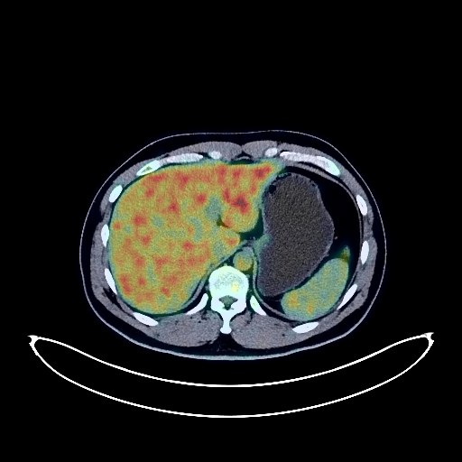
Renal Cancer PET/CT (case 983777-000001 from PETWB-REP)
Whole-body 18F-FDG PET/CT scan in a patient with Renal Cancer taken from the PETWB-REP dataset. The following English report (translated from original Chinese) is taken verbatim from the public dataset and has not been modified or otherwise checked for accuracy (see the end for citation). Impression a. Thickening of the rectal wall in the middle and upper segments, with increased FDG metabolism, suggestive of rectal cancer. Multiple lymph node metastases in the perirectal space, bilateral common iliac vessels, and retroperitoneum. b. Continuous increased FDG metabolism in the remaining colon and rectum, suggestive of inflammation or physiological changes; endoscopic follow-up is recommended. Emphysema and bullae in the upper lobes of both lungs, old tuberculous lesions in the right upper lobe, bronchiectasis in the left upper lobe, multiple chronic inflammatory micronodules in both lungs, and scattered post-inflammatory lesions in both lungs. Chronic gastritis; endoscopic follow-up is recommended. Cholestasis in the gallbladder; ultrasound follow-up is recommended. Small amount of hydrocele in both testes. Mild anterior slippage of the L4 vertebral body. Spinal osteophyte formation. L3/4, L4/5, and L5/S1 intervertebral disc bulging. Degenerative changes with inflammatory metabolism in the right L4/5 facet joint are the primary consideration; CT follow-up is recommended to rule out other possibilities. No obvious abnormalities were found on cranial imaging. Right maxillary sinusitis. This case is from PETWB-REP, a curated dataset of whole-body 18F-FDG PET/CT scans and corresponding radiology reports from 490 patients with a broad spectrum of malignancies. The data were retrospectively collected from patients who underwent clinically indicated whole-body 18F-FDG PET/CT scans at the Shanghai Universal Medical Imaging Diagnostic Center between 2021 and 2024. License: Creative Commons Attribution 4.0 International (CC BY 4.0) Citation: Xue, L., Feng, G., Wenbo, Z., Zhang, Y., Li, L., Wang, S., Peng, L., Peng, S., & Gao, X. (2026). PETWB-REP: A Multi-Cancer Whole-Body FDG PET/CT Dataset with Corresponding Radiology Reports [Data set]. Zenodo. https://doi.org/10.5281/zenodo.18670487
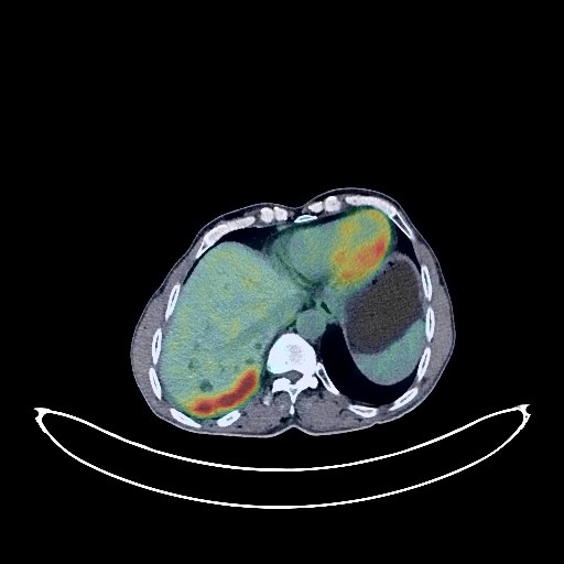
Renal Cancer PET/CT (case 983777-000004 from PETWB-REP)
Whole-body 18F-FDG PET/CT scan in a patient with Renal Cancer taken from the PETWB-REP dataset. The following English report (translated from original Chinese) is taken verbatim from the public dataset and has not been modified or otherwise checked for accuracy (see the end for citation). Impression a. Thickening of the rectal wall in the upper and middle segment, involving the adjacent sigmoid colon; increased FDG metabolism, with further increased metabolism after delay, consistent with rectal cancer. b. Metastasis to the surrounding fat spaces and presacral lymph nodes is highly probable; please correlate with clinicopathology. Multiple chronic inflammatory micronodules in both lungs; scattered post-inflammatory remnants in both lungs. Slight thickening of the pleura bilaterally. Lipoma in the subcutaneous intermuscular region of the right shoulder. Chronic gastritis; endoscopic follow-up is recommended. Cholestasis in the gallbladder; ultrasound follow-up is recommended. Left renal cyst. Spinal osteophyte formation. L3-4 vertebral endplate inflammation. L3/4 and L4/5 intervertebral disc bulge. No obvious abnormalities were seen on cranial imaging. Bilateral maxillary sinusitis. This case is from PETWB-REP, a curated dataset of whole-body 18F-FDG PET/CT scans and corresponding radiology reports from 490 patients with a broad spectrum of malignancies. The data were retrospectively collected from patients who underwent clinically indicated whole-body 18F-FDG PET/CT scans at the Shanghai Universal Medical Imaging Diagnostic Center between 2021 and 2024. License: Creative Commons Attribution 4.0 International (CC BY 4.0) Citation: Xue, L., Feng, G., Wenbo, Z., Zhang, Y., Li, L., Wang, S., Peng, L., Peng, S., & Gao, X. (2026). PETWB-REP: A Multi-Cancer Whole-Body FDG PET/CT Dataset with Corresponding Radiology Reports [Data set]. Zenodo. https://doi.org/10.5281/zenodo.18670487
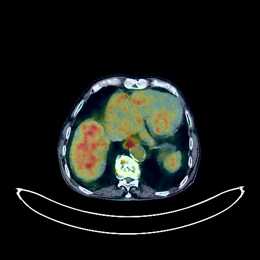
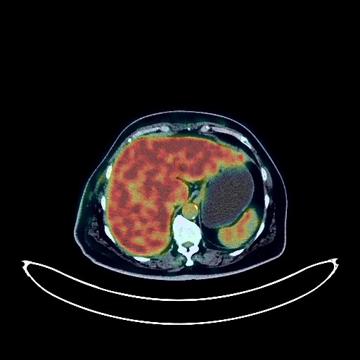
Renal Cancer PET/CT (case 983777-000006 from PETWB-REP)
Whole-body 18F-FDG PET/CT scan in a patient with Renal Cancer taken from the PETWB-REP dataset. The following English report (translated from original Chinese) is taken verbatim from the public dataset and has not been modified or otherwise checked for accuracy (see the end for citation). Impression a. Thickening of the lower descending colon near the sigmoid colon with increased FDG metabolism, consistent with colon cancer. Mesenteric lymph node metastasis. Liver metastasis. b. Increased FDG metabolism in the remaining intestinal segment, suggesting physiological changes or inflammatory lesions; chronic gastritis. Please follow up with endoscopy for the above. Multiple ground-glass nodules in both lungs, normal FDG metabolism, atypical adenomatous hyperplasia or early-stage lung cancer (larger ones) are the primary considerations; CT scan follow-up in 3 months is recommended. Chronic inflammatory micronodules (solid) in both lungs. Anemia. Nodular goiter, please follow up with ultrasound. Contrast agent residue or cholestasis in the gallbladder. Contrast agent residue in the intestines. Multiple uterine fibroids, Nabothian cysts in the cervix, please follow up with ultrasound. Small amount of pelvic effusion. Mild osteophyte formation in the spine. No significant abnormalities in FDG metabolism in the brain. This case is from PETWB-REP, a curated dataset of whole-body 18F-FDG PET/CT scans and corresponding radiology reports from 490 patients with a broad spectrum of malignancies. The data were retrospectively collected from patients who underwent clinically indicated whole-body 18F-FDG PET/CT scans at the Shanghai Universal Medical Imaging Diagnostic Center between 2021 and 2024. License: Creative Commons Attribution 4.0 International (CC BY 4.0) Citation: Xue, L., Feng, G., Wenbo, Z., Zhang, Y., Li, L., Wang, S., Peng, L., Peng, S., & Gao, X. (2026). PETWB-REP: A Multi-Cancer Whole-Body FDG PET/CT Dataset with Corresponding Radiology Reports [Data set]. Zenodo. https://doi.org/10.5281/zenodo.18670487
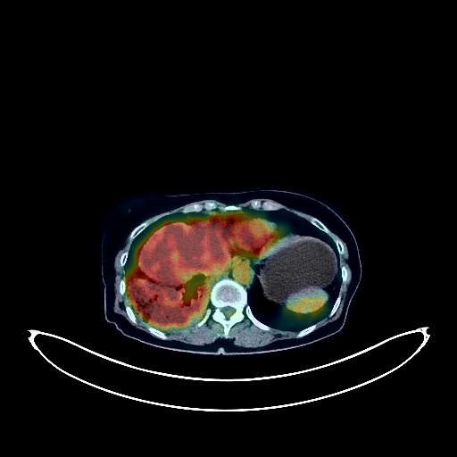
Renal Cancer PET/CT (case 983777-000007 from PETWB-REP)
Whole-body 18F-FDG PET/CT scan in a patient with Renal Cancer taken from the PETWB-REP dataset. The following English report (translated from original Chinese) is taken verbatim from the public dataset and has not been modified or otherwise checked for accuracy (see the end for citation). Impression Irregular thickening of the mid-rectal wall with elevated FDG metabolism, suggestive of rectal cancer based on medical history. Reactive hyperplasia of small presacral lymph nodes, close observation recommended to rule out metastasis. Small amount of pelvic effusion. Chronic inflammatory micronodule in the upper lobe of the left lung, follow-up CT recommended. A few post-inflammatory lesions in both lungs. Mild anemia, slight arteriosclerosis in some arteries. Small nodule in the right breast, FDG metabolism normal, suggestive of hyperplastic nodule or fibroadenoma, follow-up ultrasound recommended. Small cyst in the left lateral lobe of the liver, hemangioma in the left medial lobe and right anterior lobe of the liver is the first consideration, please combine with MRI. Uterine fibroid. Chronic inflammatory changes in part of the gastric wall, please combine with endoscopy. Degenerative changes in the spine. L4/5 and L5/S1 intervertebral disc bulge. Low-density nodule in the left lobe of the thyroid, FDG metabolism normal, suggestive of adenomatous nodule, please combine with ultrasound. No obvious abnormalities seen on cranial scintigraphy. Bilateral chronic ethmoid sinusitis. This case is from PETWB-REP, a curated dataset of whole-body 18F-FDG PET/CT scans and corresponding radiology reports from 490 patients with a broad spectrum of malignancies. The data were retrospectively collected from patients who underwent clinically indicated whole-body 18F-FDG PET/CT scans at the Shanghai Universal Medical Imaging Diagnostic Center between 2021 and 2024. License: Creative Commons Attribution 4.0 International (CC BY 4.0) Citation: Xue, L., Feng, G., Wenbo, Z., Zhang, Y., Li, L., Wang, S., Peng, L., Peng, S., & Gao, X. (2026). PETWB-REP: A Multi-Cancer Whole-Body FDG PET/CT Dataset with Corresponding Radiology Reports [Data set]. Zenodo. https://doi.org/10.5281/zenodo.18670487
Lymphoma PET/CT (case 983824-000001 from PETWB-REP)
Whole-body 18F-FDG PET/CT scan in a patient with Lymphoma taken from the PETWB-REP dataset. The following English report (translated from original Chinese) is taken verbatim from the public dataset and has not been modified or otherwise checked for accuracy (see the end for citation). Impression Following comprehensive treatment for lymphoma, comparing PET/CT images from our center on December 25, 2021: a. Postoperative gastric lesser curvature lymphoma surgery showed slight thickening of the gastric wall in the surgical area with mild FDG uptake, suggesting postoperative changes; the soft tissue nodules in the left posteroinferior mediastinum (medial to the descending aorta at T8 level) decreased in size, with reduced FDG metabolism; the right inguinal lymph nodes also decreased in size, with reduced FDG metabolism. These findings suggest effective lymphoma treatment. b. Multiple small lymph nodes throughout the body (see description for details) showed no significant increase in FDG metabolism, remaining similar to previous findings, suggesting suppressed lesion activity or possibly reactive proliferative lymph nodes after treatment. c. Patchy and strip-like lesions in both lungs with increased FDG metabolism, some adjacent pleural thickening and adhesions, and some newly added lesions suggest a high probability of inflammatory lesions. Please follow up with CT scans to rule out other possibilities. Multiple thin-walled cystic lesions in both lungs, scattered post-inflammatory remnants (including calcifications) in both lungs, roughly similar to previous findings. A small amount of effusion in the pericardial recesses and at their bases. Partial arteriosclerosis. Bilateral breast hyperplasia. Multiple solid lesions in the liver, roughly similar to previous findings, suggestive of hemangioma; enhanced MRI follow-up is recommended. A small nodule in the left adrenal region, roughly similar to previous findings, highly suggestive of adenoma. Chronic inflammatory changes or physiological uptake in some intestinal segments; endoscopic follow-up is recommended. Duodenal diverticulum. Hemorrhoidal changes. Scoliosis with degenerative changes. Post-radiotherapy changes in some thoracic spine segments. Mild L2/3 intervertebral disc bulge. Lipoma and calcifications in the quadrigeminal cistern. A small amount of inflammation in the right maxillary sinus. Thyroid gland density is uneven; FDG metabolism is normal; ultrasound follow-up is recommended. This case is from PETWB-REP, a curated dataset of whole-body 18F-FDG PET/CT scans and corresponding radiology reports from 490 patients with a broad spectrum of malignancies. The data were retrospectively collected from patients who underwent clinically indicated whole-body 18F-FDG PET/CT scans at the Shanghai Universal Medical Imaging Diagnostic Center between 2021 and 2024. License: Creative Commons Attribution 4.0 International (CC BY 4.0) Citation: Xue, L., Feng, G., Wenbo, Z., Zhang, Y., Li, L., Wang, S., Peng, L., Peng, S., & Gao, X. (2026). PETWB-REP: A Multi-Cancer Whole-Body FDG PET/CT Dataset with Corresponding Radiology Reports [Data set]. Zenodo. https://doi.org/10.5281/zenodo.18670487
Lung Cancer PET/CT (case 983824-000002 from PETWB-REP)
Whole-body 18F-FDG PET/CT scan in a patient with Lung Cancer taken from the PETWB-REP dataset. The following English report (translated from original Chinese) is taken verbatim from the public dataset and has not been modified or otherwise checked for accuracy (see the end for citation). Impression a. A nodule at the left apex of the lung, highly suggestive of primary lung cancer; please correlate with clinicopathology. Enlarged hilar and mediastinal lymph nodes bilaterally with mildly elevated FDG metabolism suggest metastasis; follow-up examination after treatment is recommended. b. Left pleural effusion, multiple thickenings of the left pleura with elevated FDG metabolism suggest multiple pleural metastases. c. Destruction of the left portion of the T10 vertebral body and the left 10th and 11th posterior ribs with soft tissue mass, elevated FDG metabolism, suggest metastasis. Scattered emphysema and bullae in both lungs; cystic dilatation of some bronchi in the right middle and lower lobes. Mixed ground-glass nodules in the posterior segment of the right lower lobe and the anterior-medial basal segment of the left lower lobe, with no elevated FDG metabolism, suggestive of atypical adenomatous hyperplasia or chronic inflammatory nodules; multiple chronic inflammations and old lesions in both lungs (possible old pulmonary tuberculosis in the right upper lobe). Localized thickening of the pleura in both oblique fissures. Calcification of some arterial walls (including coronary arteries). Localized soft tissue nodules in the mid-sigmoid colon with increased FDG metabolism, suggestive of a polyp with potential malignancy; please confirm with colonoscopy pathology. Inflammatory changes in the lower sigmoid colon and rectum, reactive hyperplasia of lymph nodes at the root of the mesocolic colon. Small cyst in the left lobe of the liver. Prostatic calcification. Calcification of the tunica vaginalis on the left side of the testis, bilateral hydrocele. Spinal osteophyte formation. Lacunar ischemic foci in the bilateral basal ganglia. Age-related brain changes. Chronic inflammation of the nasopharynx. Minor inflammation of the right maxillary sinus. This case is from PETWB-REP, a curated dataset of whole-body 18F-FDG PET/CT scans and corresponding radiology reports from 490 patients with a broad spectrum of malignancies. The data were retrospectively collected from patients who underwent clinically indicated whole-body 18F-FDG PET/CT scans at the Shanghai Universal Medical Imaging Diagnostic Center between 2021 and 2024. License: Creative Commons Attribution 4.0 International (CC BY 4.0) Citation: Xue, L., Feng, G., Wenbo, Z., Zhang, Y., Li, L., Wang, S., Peng, L., Peng, S., & Gao, X. (2026). PETWB-REP: A Multi-Cancer Whole-Body FDG PET/CT Dataset with Corresponding Radiology Reports [Data set]. Zenodo. https://doi.org/10.5281/zenodo.18670487
Pancreatic Cancer PET/CT (case 983824-000003 from PETWB-REP)
Whole-body 18F-FDG PET/CT scan in a patient with Pancreatic Cancer taken from the PETWB-REP dataset. The following English report (translated from original Chinese) is taken verbatim from the public dataset and has not been modified or otherwise checked for accuracy (see the end for citation). Impression a. After chemotherapy for pancreatic cancer, a soft tissue mass in the pancreatic body with increased FDG metabolism suggests continued tumor activity; peripancreatic lymph nodes show slightly increased FDG metabolism. It is recommended to compare these findings with pre-treatment imaging data and follow up. b. After radiofrequency ablation of liver metastases, multiple low-density nodules and masses were observed in the liver. The lesion in the right lobe showed loss of FDG uptake, but no significant increase in FDG metabolism, suggesting suppressed tumor activity after treatment. c. No space-occupying lesions were observed in either kidney, and FDG metabolism was normal. Enhanced MRI follow-up is recommended. a. Ground-glass nodules in the apical segment of the right upper lobe, the posterior basal segment of the left lower lobe, and the lateral basal segment of the right lower lobe, with normal FDG metabolism, suggest inflammatory nodules or atypical adenomatous hyperplasia. Annual HRCT follow-up is recommended. b. Calcification in the right upper lobe, and a few post-inflammatory remnants in both lungs. Calcification of some arterial walls (including coronary arteries). Multiple liver cysts. Calcification in the right inguinal canal. Degenerative changes in the spine, with L4/5 and L5/S1 intervertebral disc bulges. Post-fracture changes of the left 7th-10th ribs. No obvious abnormalities were found on cranial scintigraphy. This case is from PETWB-REP, a curated dataset of whole-body 18F-FDG PET/CT scans and corresponding radiology reports from 490 patients with a broad spectrum of malignancies. The data were retrospectively collected from patients who underwent clinically indicated whole-body 18F-FDG PET/CT scans at the Shanghai Universal Medical Imaging Diagnostic Center between 2021 and 2024. License: Creative Commons Attribution 4.0 International (CC BY 4.0) Citation: Xue, L., Feng, G., Wenbo, Z., Zhang, Y., Li, L., Wang, S., Peng, L., Peng, S., & Gao, X. (2026). PETWB-REP: A Multi-Cancer Whole-Body FDG PET/CT Dataset with Corresponding Radiology Reports [Data set]. Zenodo. https://doi.org/10.5281/zenodo.18670487
Lung Cancer PET/CT (case 983824-000004 from PETWB-REP)
Whole-body 18F-FDG PET/CT scan in a patient with Lung Cancer taken from the PETWB-REP dataset. The following English report (translated from original Chinese) is taken verbatim from the public dataset and has not been modified or otherwise checked for accuracy (see the end for citation). Impression a. Irregular nodule in the apical segment of the right upper lobe, with elevated FDG metabolism, suggestive of peripheral lung cancer; further clinical pathology is recommended. b. Several miliary chronic inflammatory nodules in both lungs. A few old lesions in both lungs. Emphysema in both lungs. Calcification of some arterial walls (including coronary arteries). Cystic mass in the head and neck of the pancreas, cystadenoma to be ruled out; further examination with contrast-enhanced MRI is recommended. Multiple cysts in the liver. Bilateral hydrocele. Degenerative changes in the spine. L4/5 and L5/S1 intervertebral disc bulge. Fat deposition in the right hip bone. Age-related brain changes. This case is from PETWB-REP, a curated dataset of whole-body 18F-FDG PET/CT scans and corresponding radiology reports from 490 patients with a broad spectrum of malignancies. The data were retrospectively collected from patients who underwent clinically indicated whole-body 18F-FDG PET/CT scans at the Shanghai Universal Medical Imaging Diagnostic Center between 2021 and 2024. License: Creative Commons Attribution 4.0 International (CC BY 4.0) Citation: Xue, L., Feng, G., Wenbo, Z., Zhang, Y., Li, L., Wang, S., Peng, L., Peng, S., & Gao, X. (2026). PETWB-REP: A Multi-Cancer Whole-Body FDG PET/CT Dataset with Corresponding Radiology Reports [Data set]. Zenodo. https://doi.org/10.5281/zenodo.18670487
Colon Cancer PET/CT (case 983824-000005 from PETWB-REP)
Whole-body 18F-FDG PET/CT scan in a patient with Colon Cancer taken from the PETWB-REP dataset. The following English report (translated from original Chinese) is taken verbatim from the public dataset and has not been modified or otherwise checked for accuracy (see the end for citation). Impression Thickening of the ascending colon near the ileocecal junction with increased FDG uptake, thickening of the surrounding mesentery with several lymph nodes visible. Delayed scanning showed no significant change in the lesion near the ileocecal junction, but FDG uptake values in all lesions remained increased, suggesting possible colon cancer with surrounding mesenteric lymph node metastasis. A follow-up colonoscopy is recommended to rule out other possibilities; inflammatory uptake in the colon is also possible. Liver cirrhosis findings, gallstones in the gallbladder neck; no space-occupying lesions or foci of increased FDG uptake were observed in the liver, pancreas, or biliary system. Clinical and MRI findings are recommended. Thickening of the gastric wall with mildly increased FDG uptake is suggested, possibly due to inflammatory uptake. A follow-up gastroscopy is recommended. Multiple solid nodules in both lungs with increased FDG uptake are suggested, possibly due to inflammatory nodules. Clinical and old film comparison and follow-up CT scan are recommended. Interstitial pneumonia in the lower lobes of both lungs. Slight pleural thickening bilaterally. Reactive hyperplasia of hilar and mediastinal lymph nodes bilaterally. Calcification of some arterial walls (including coronary arteries). Bilateral renal atrophy, small renal stone in the left kidney. Benign prostatic hyperplasia with calcification, uneven FDG metabolism; please follow up with PSA levels. Bilateral hydrocele. Spinal degenerative changes. L4/5 and L5/S1 intervertebral disc bulge. Bilateral subcutaneous calcifications in the buttocks. Bilateral basal ganglia ischemia. Age-related brain changes. Bilateral ethmoid sinusitis and right maxillary sinusitis. This case is from PETWB-REP, a curated dataset of whole-body 18F-FDG PET/CT scans and corresponding radiology reports from 490 patients with a broad spectrum of malignancies. The data were retrospectively collected from patients who underwent clinically indicated whole-body 18F-FDG PET/CT scans at the Shanghai Universal Medical Imaging Diagnostic Center between 2021 and 2024. License: Creative Commons Attribution 4.0 International (CC BY 4.0) Citation: Xue, L., Feng, G., Wenbo, Z., Zhang, Y., Li, L., Wang, S., Peng, L., Peng, S., & Gao, X. (2026). PETWB-REP: A Multi-Cancer Whole-Body FDG PET/CT Dataset with Corresponding Radiology Reports [Data set]. Zenodo. https://doi.org/10.5281/zenodo.18670487
Lung Cancer PET/CT (case 983824-000007 from PETWB-REP)
Whole-body 18F-FDG PET/CT scan in a patient with Lung Cancer taken from the PETWB-REP dataset. The following English report (translated from original Chinese) is taken verbatim from the public dataset and has not been modified or otherwise checked for accuracy (see the end for citation). Impression a. Soft tissue mass in the left upper lobe of the lung with increased FDG metabolism, consistent with lung cancer based on pathology. b. Solid nodule in the posterior segment of the left upper lobe with increased FDG metabolism, suggestive of metastasis. c. Left hilar lymph node metastasis. Multiple bone metastases throughout the body (see description for details). d. Multiple small solid nodules in the remaining lungs, with no significant FDG uptake, highly suggestive of chronic inflammatory nodules; follow-up is recommended. Interstitial fibrosis in both lungs with scattered chronic inflammation and remnants. Bilateral pleural thickening. Bilateral incomplete breast regression; follow-up with ultrasound is recommended. Postoperative changes in the pancreatic mass; no abnormalities were found in FDG metabolism at the surgical site; follow-up is recommended. Highly suggestive of a liver cyst. Thickening of the left perirenal fascia, possible complex cyst in the left kidney; calcification of the left renal papillae; follow-up with ultrasound is recommended. a. Post-gastric surgery changes: increased FDG metabolism in part of the anastomotic wall, possibly due to physiological uptake or chronic inflammation. Follow-up with clinical findings and gastroscopy is recommended. b. Increased FDG metabolism in part of the intestinal tract, possibly due to physiological uptake or chronic inflammation. Further colonoscopy is recommended to rule out other possibilities. Osteoporosis. Spinal degeneration. L2/3, L3/4, L4/5 disc bulging, L5/S1 disc herniation. Right external oblique muscle lipoma. Bilateral deep lacunar infarcts, age-related brain abnormalities. MRI is recommended. Unevenly decreased density in the left and right lobes of the thyroid gland, with no abnormalities in FDG metabolism. Follow-up with ultrasound is recommended. This case is from PETWB-REP, a curated dataset of whole-body 18F-FDG PET/CT scans and corresponding radiology reports from 490 patients with a broad spectrum of malignancies. The data were retrospectively collected from patients who underwent clinically indicated whole-body 18F-FDG PET/CT scans at the Shanghai Universal Medical Imaging Diagnostic Center between 2021 and 2024. License: Creative Commons Attribution 4.0 International (CC BY 4.0) Citation: Xue, L., Feng, G., Wenbo, Z., Zhang, Y., Li, L., Wang, S., Peng, L., Peng, S., & Gao, X. (2026). PETWB-REP: A Multi-Cancer Whole-Body FDG PET/CT Dataset with Corresponding Radiology Reports [Data set]. Zenodo. https://doi.org/10.5281/zenodo.18670487
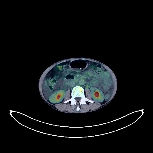
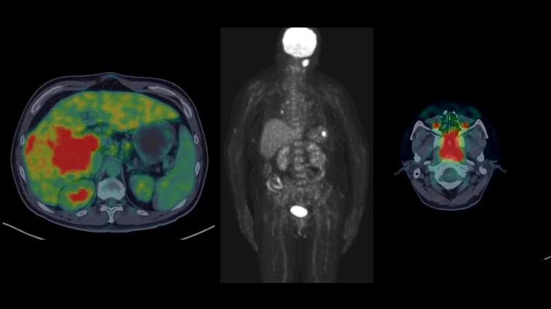
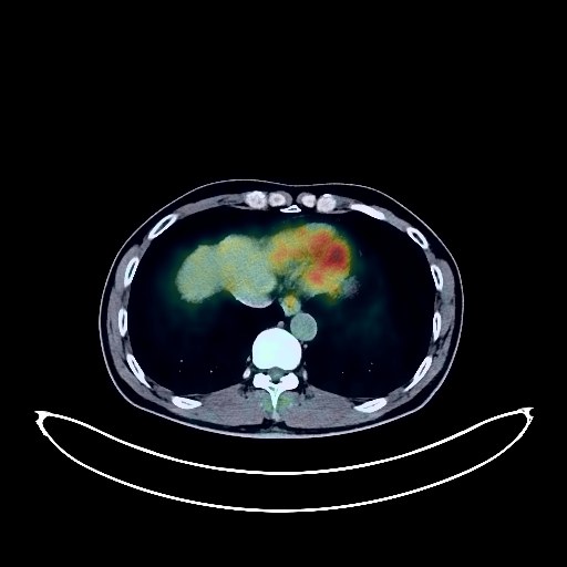
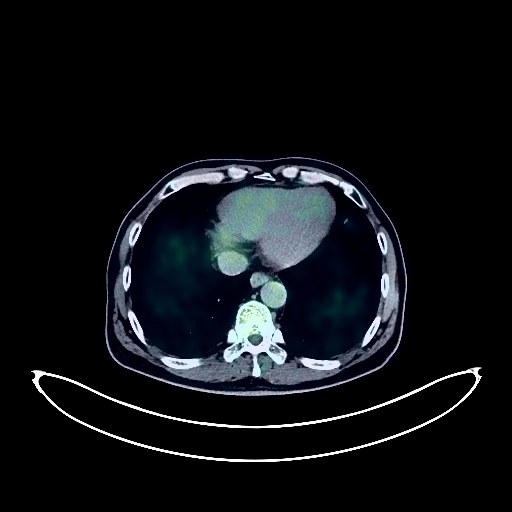
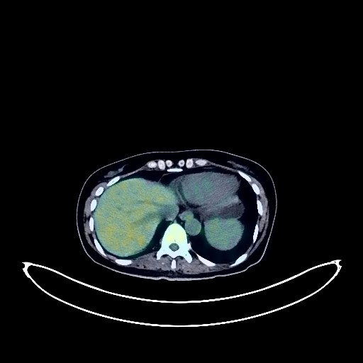
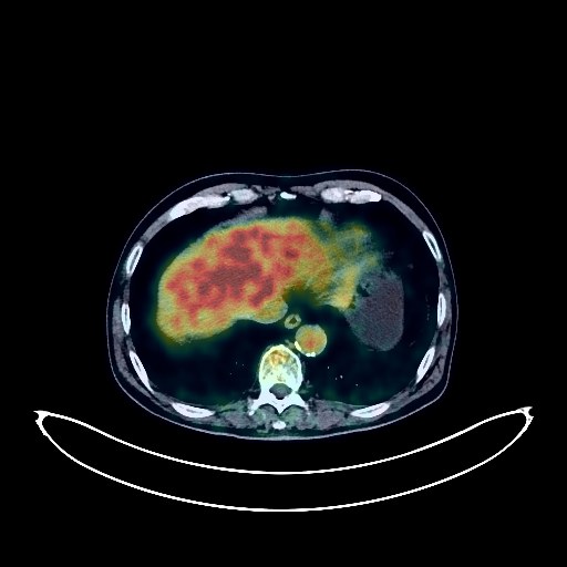
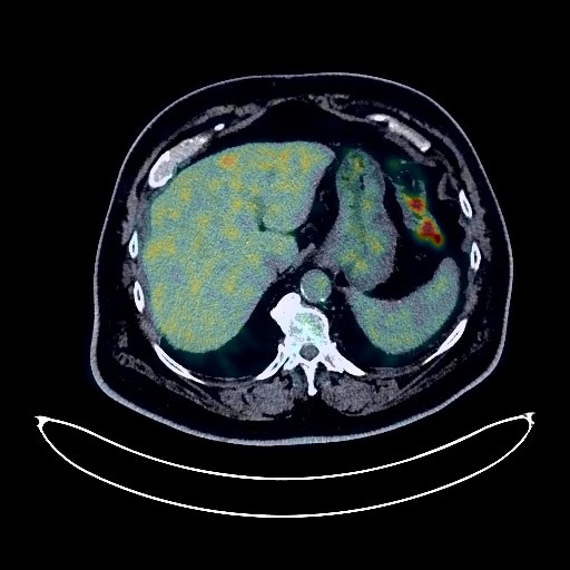
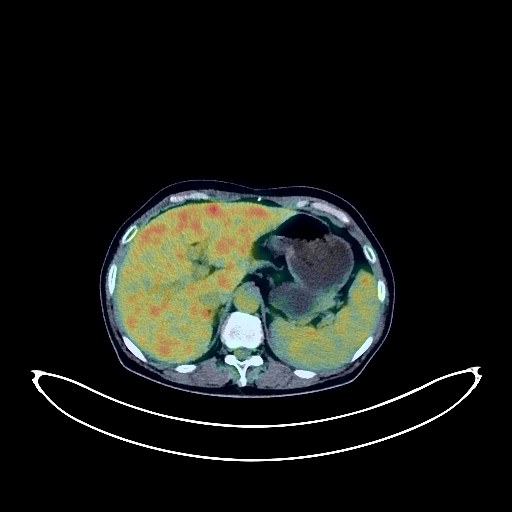
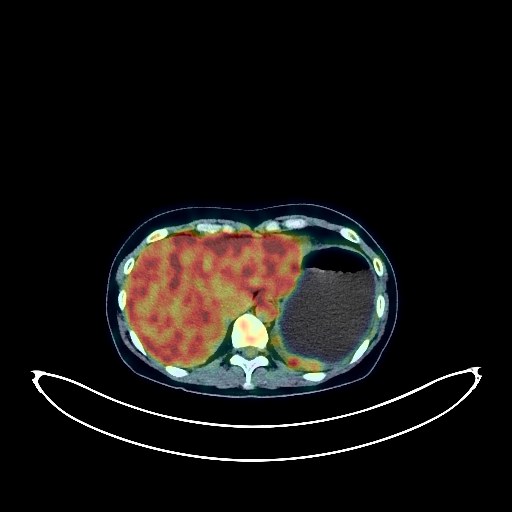
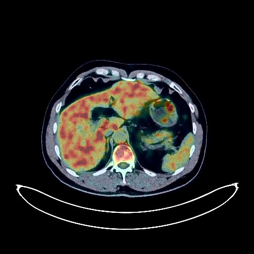
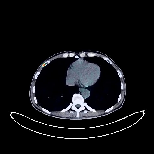
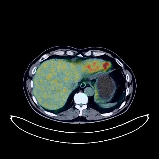
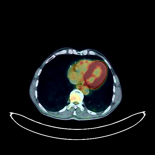
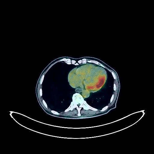
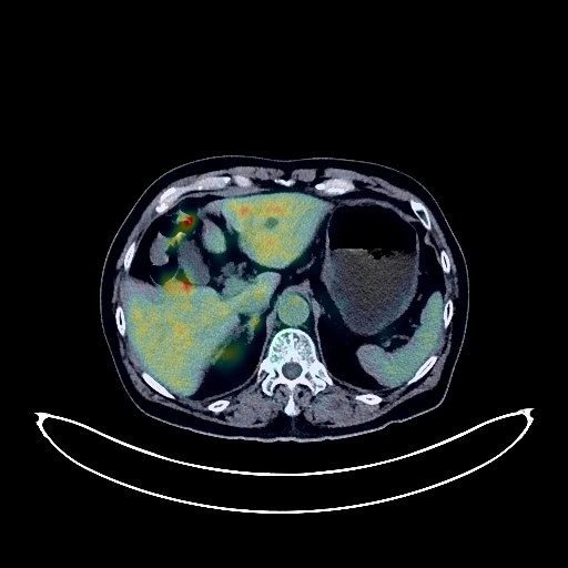
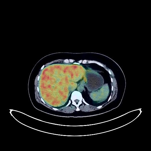
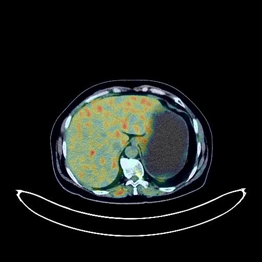
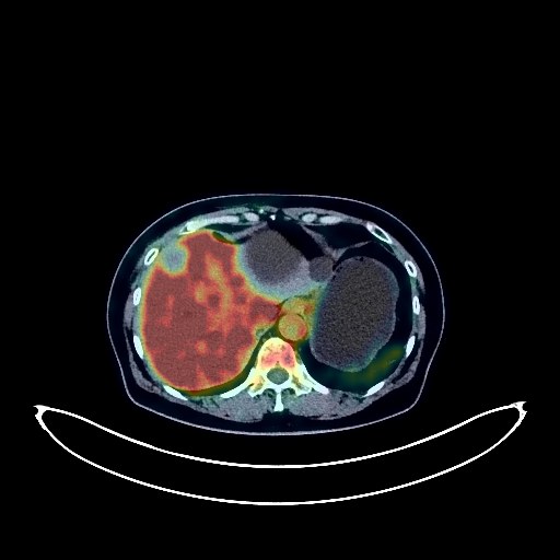
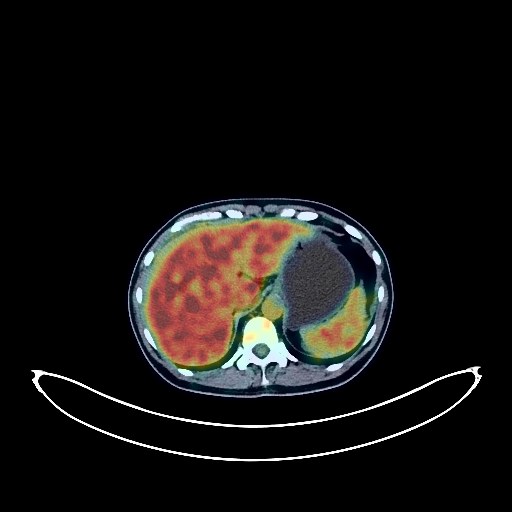
Liver Cancer PET/CT (case 983824-000008 from PETWB-REP)
Whole-body 18F-FDG PET/CT scan in a patient with Liver Cancer taken from the PETWB-REP dataset. The following English report (translated from original Chinese) is taken verbatim from the public dataset and has not been modified or otherwise checked for accuracy (see the end for citation). Impression a. Multiple space-occupying lesions in the liver, with increased FDG metabolism, suggestive of malignancy, most likely primary liver cancer. b. Multiple lymph node metastases in the hilum and retroperitoneum. Diffuse metastases in both lungs. Extensive bone metastases throughout the body (pathological fracture of the right 3rd posterior rib). c. Liver cirrhosis; splenomegaly. Large amounts of fluid in the abdomen and pelvis. A few chronic lesions and old lesions in both lungs. Degenerative changes in the spine. L4/5 and L5/S1 intervertebral disc bulges. Mild age-related brain changes. Sclerotic mastoid process on the left side. This case is from PETWB-REP, a curated dataset of whole-body 18F-FDG PET/CT scans and corresponding radiology reports from 490 patients with a broad spectrum of malignancies. The data were retrospectively collected from patients who underwent clinically indicated whole-body 18F-FDG PET/CT scans at the Shanghai Universal Medical Imaging Diagnostic Center between 2021 and 2024. License: Creative Commons Attribution 4.0 International (CC BY 4.0) Citation: Xue, L., Feng, G., Wenbo, Z., Zhang, Y., Li, L., Wang, S., Peng, L., Peng, S., & Gao, X. (2026). PETWB-REP: A Multi-Cancer Whole-Body FDG PET/CT Dataset with Corresponding Radiology Reports [Data set]. Zenodo. https://doi.org/10.5281/zenodo.18670487
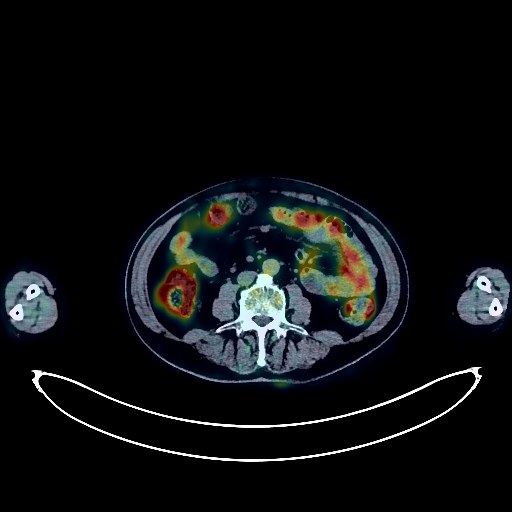
Colon Cancer PET/CT (case 983824-000009 from PETWB-REP)
Whole-body 18F-FDG PET/CT scan in a patient with Colon Cancer taken from the PETWB-REP dataset. The following English report (translated from original Chinese) is taken verbatim from the public dataset and has not been modified or otherwise checked for accuracy (see the end for citation). Impression a. Multiple peritoneal metastases in the abdominopelvic cavity (including perihepatic, hilar, and perisplenic areas); right lower abdominal wall metastasis; left inguinal region metastasis. b. A mass in the sigmoid colon with increased FDG uptake, suggestive of malignancy, most likely colon cancer; please follow up with colonoscopy. Liver cirrhosis. Calcification in the right lobe of the liver. A few calcifications in the spleen. Scattered chronic inflammatory nodules in both lungs; please follow up with CT scan. Interstitial lung changes with emphysema in both lungs. Chronic inflammation and sequelae in both lungs. Reactive hyperplasia of bilateral supraclavicular and mediastinal lymph nodes. Cardiac enlargement. Calcification of some arterial walls (including coronary arteries); please follow up with coronary CTA. Highly probable inflammatory uptake in the middle and lower esophagus; please follow up with gastroscopy. Mild renal atrophy in both kidneys. Left renal cyst. Right renal calculus. Calcification and hydrocele in the right testicular tunica vaginalis. Osteoporosis. Compression changes in the T10 vertebral body. Spinal degeneration. Right hip periarthritis. A few ischemic foci deep in the brain, age-related brain changes. This case is from PETWB-REP, a curated dataset of whole-body 18F-FDG PET/CT scans and corresponding radiology reports from 490 patients with a broad spectrum of malignancies. The data were retrospectively collected from patients who underwent clinically indicated whole-body 18F-FDG PET/CT scans at the Shanghai Universal Medical Imaging Diagnostic Center between 2021 and 2024. License: Creative Commons Attribution 4.0 International (CC BY 4.0) Citation: Xue, L., Feng, G., Wenbo, Z., Zhang, Y., Li, L., Wang, S., Peng, L., Peng, S., & Gao, X. (2026). PETWB-REP: A Multi-Cancer Whole-Body FDG PET/CT Dataset with Corresponding Radiology Reports [Data set]. Zenodo. https://doi.org/10.5281/zenodo.18670487
Colon Cancer PET/CT (case 983824-000010 from PETWB-REP)
Whole-body 18F-FDG PET/CT scan in a patient with Colon Cancer taken from the PETWB-REP dataset. The following English report (translated from original Chinese) is taken verbatim from the public dataset and has not been modified or otherwise checked for accuracy (see the end for citation). Impression a. A cystic-solid mass in the pelvic cavity with increased FDG metabolism at the periphery, suggesting a high probability of neoplastic lesions (metastasis is more likely than primary malignant ovarian tumors). Inflammatory lesions (tuberculosis) need to be ruled out. Please combine laboratory tests and clinicopathological examination. b. Pelvic effusion, extensive thickening and blurring of the perihepatic and splenic capsule and peritoneum in the abdominopelvic cavity, with multiple small lymph nodes visible in the mesentery, and increased FDG metabolism, suggesting extensive involvement of the pelvic and abdominal cavities. c. No obvious abnormal FDG metabolism was observed in the pancreas and biliary system (further CA19-9 and enhanced MRI follow-up are recommended). Scattered ground-glass nodules in the lateral basal segment of the left lower lobe and the apical segment of the right upper lobe, with no increased FDG metabolism, suggestive of atypical adenomatous hyperplasia or chronic inflammatory nodules. A small chronic inflammatory nodule in the posterior segment of the right upper lobe, please follow up with CT. Small amount of pleural effusion in the left side. Bilateral solid breast nodules, with no increased FDG metabolism, suggestive of fibroadenomas, please combine with ultrasound follow-up. Small hepatic cysts. Accessory spleen. Increased FDG metabolism in the rectum and anal canal, likely due to inflammatory uptake; please confirm clinical findings and repeat colonoscopy to rule out the possibility of a low-lying tumor. Partial vertebral osteophyte formation. Nuchal ligament calcification. No obvious abnormalities found on cranial scintigraphy. This case is from PETWB-REP, a curated dataset of whole-body 18F-FDG PET/CT scans and corresponding radiology reports from 490 patients with a broad spectrum of malignancies. The data were retrospectively collected from patients who underwent clinically indicated whole-body 18F-FDG PET/CT scans at the Shanghai Universal Medical Imaging Diagnostic Center between 2021 and 2024. License: Creative Commons Attribution 4.0 International (CC BY 4.0) Citation: Xue, L., Feng, G., Wenbo, Z., Zhang, Y., Li, L., Wang, S., Peng, L., Peng, S., & Gao, X. (2026). PETWB-REP: A Multi-Cancer Whole-Body FDG PET/CT Dataset with Corresponding Radiology Reports [Data set]. Zenodo. https://doi.org/10.5281/zenodo.18670487
Renal Cancer PET/CT (case 983824-000011 from PETWB-REP)
Whole-body 18F-FDG PET/CT scan in a patient with Renal Cancer taken from the PETWB-REP dataset. The following English report (translated from original Chinese) is taken verbatim from the public dataset and has not been modified or otherwise checked for accuracy (see the end for citation). Impression a. A slightly low-density soft tissue lesion in the left kidney, with background uptake on FDG, suggestive of a renal space-occupying lesion, possibly malignant. Please confirm with contrast-enhanced MRI and clinicopathological examination. Small renal stone in the left kidney. Cyst in the right kidney. b. A soft tissue lesion in the right anterior lobe of the liver, with background uptake on FDG, highly suggestive of a neoplastic lesion. Chronic inflammatory nodules in the posterior segment of the left upper lobe and the apical segment of the right upper lobe. Please confirm with CT follow-up. Soft tissue nodules in the anterior mediastinum, with absent FDG metabolism, suggestive of a thymic cyst or thymoma. Please confirm with CT follow-up. Calcification of some arterial walls. Benign prostatic hyperplasia. Degenerative changes in the spine. Wedge-shaped L2 vertebral body. Intervertebral disc bulging at L3/4, L4/5, and L5/S1. No obvious abnormalities were found on cranial scintigraphy. This case is from PETWB-REP, a curated dataset of whole-body 18F-FDG PET/CT scans and corresponding radiology reports from 490 patients with a broad spectrum of malignancies. The data were retrospectively collected from patients who underwent clinically indicated whole-body 18F-FDG PET/CT scans at the Shanghai Universal Medical Imaging Diagnostic Center between 2021 and 2024. License: Creative Commons Attribution 4.0 International (CC BY 4.0) Citation: Xue, L., Feng, G., Wenbo, Z., Zhang, Y., Li, L., Wang, S., Peng, L., Peng, S., & Gao, X. (2026). PETWB-REP: A Multi-Cancer Whole-Body FDG PET/CT Dataset with Corresponding Radiology Reports [Data set]. Zenodo. https://doi.org/10.5281/zenodo.18670487
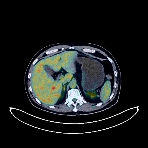
Pancreatic Cancer PET/CT (case 983824-000012 from PETWB-REP)
Whole-body 18F-FDG PET/CT scan in a patient with Pancreatic Cancer taken from the PETWB-REP dataset. The following English report (translated from original Chinese) is taken verbatim from the public dataset and has not been modified or otherwise checked for accuracy (see the end for citation). Impression a. A slightly low-density mass in the tail of the pancreas with increased FDG metabolism, poorly demarcated from the splenic hilum, highly suggestive of pancreatic cancer; please correlate with clinicopathology. b. Multiple liver metastases. Metastases in the hilar and retroperitoneal lymph nodes. Metastases in the right 3rd and 6th ribs, left T8 vertebral body, left iliac bone, and left pubic bone. Gallbladder wall thickening, soft tissue lesions within the gallbladder with increased FDG metabolism, suggestive of gallbladder adenomyosis; local malignancy to be ruled out; please correlate with pathology. Multiple liver cysts. Small renal cysts. Scattered chronic inflammatory nodules in both lungs. Emphysema in both lungs. Calcification in the right lung. Calcification of some arterial walls. Physiological or inflammatory uptake in some intestinal segments; please correlate with colonoscopy follow-up. Benign prostatic hyperplasia with calcification. Some vertebral osteophytes. Some thoracic intervertebral disc effusion. Age-related brain changes. This case is from PETWB-REP, a curated dataset of whole-body 18F-FDG PET/CT scans and corresponding radiology reports from 490 patients with a broad spectrum of malignancies. The data were retrospectively collected from patients who underwent clinically indicated whole-body 18F-FDG PET/CT scans at the Shanghai Universal Medical Imaging Diagnostic Center between 2021 and 2024. License: Creative Commons Attribution 4.0 International (CC BY 4.0) Citation: Xue, L., Feng, G., Wenbo, Z., Zhang, Y., Li, L., Wang, S., Peng, L., Peng, S., & Gao, X. (2026). PETWB-REP: A Multi-Cancer Whole-Body FDG PET/CT Dataset with Corresponding Radiology Reports [Data set]. Zenodo. https://doi.org/10.5281/zenodo.18670487
Prostate Cancer PET/CT (case 983824-000013 from PETWB-REP)
Whole-body 18F-FDG PET/CT scan in a patient with Prostate Cancer taken from the PETWB-REP dataset. The following English report (translated from original Chinese) is taken verbatim from the public dataset and has not been modified or otherwise checked for accuracy (see the end for citation). Impression a. Prostatic mass with elevated FDG metabolism, consistent with prostate cancer; multiple lymph node metastases bilaterally to the iliac vessels, retroperitoneum, and bilateral posterior diaphragmatic crura. b. Bone metastases to the right scapula, left transverse process of T11, and right side of L2 vertebral body. Thickening and roughening of the right bladder wall, with normal FDG metabolism, suggestive of chronic inflammation; further specialist examination is required. Chronic inflammatory nodules in both lungs. Paraseptal emphysema in the right upper lobe, with a few post-inflammatory lesions in both lungs. Pleural thickening bilaterally. Reactive hyperplasia of mediastinal lymph nodes. Mild anemia, with partial calcification of arterial walls (including coronary arteries). Bilateral renal cysts, with angiomyolipoma of the right upper pole of the kidney to be ruled out. Bilateral inguinal hernias are possible. Small amount of hydrocele in both testes. Chronic gastritis. Degenerative changes in the spine, with L4/5 and L5/S1 intervertebral disc bulges. Left hip periarthritis. Age-related brain abnormalities, deep lacunar infarcts; MRI follow-up recommended. Chronic inflammation of the left maxillary sinus. Right vocal cord laxity. This case is from PETWB-REP, a curated dataset of whole-body 18F-FDG PET/CT scans and corresponding radiology reports from 490 patients with a broad spectrum of malignancies. The data were retrospectively collected from patients who underwent clinically indicated whole-body 18F-FDG PET/CT scans at the Shanghai Universal Medical Imaging Diagnostic Center between 2021 and 2024. License: Creative Commons Attribution 4.0 International (CC BY 4.0) Citation: Xue, L., Feng, G., Wenbo, Z., Zhang, Y., Li, L., Wang, S., Peng, L., Peng, S., & Gao, X. (2026). PETWB-REP: A Multi-Cancer Whole-Body FDG PET/CT Dataset with Corresponding Radiology Reports [Data set]. Zenodo. https://doi.org/10.5281/zenodo.18670487
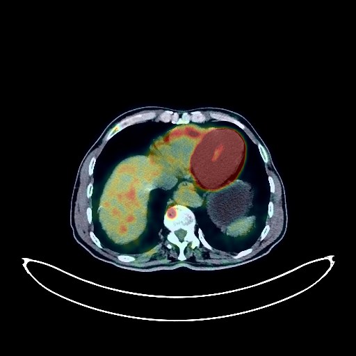
Lymphoma PET/CT (case 983824-000014 from PETWB-REP)
Whole-body 18F-FDG PET/CT scan in a patient with Lymphoma taken from the PETWB-REP dataset. The following English report (translated from original Chinese) is taken verbatim from the public dataset and has not been modified or otherwise checked for accuracy (see the end for citation). Impression a. After treatment for splenic lymphoma, no obvious space-occupying lesion was found in the spleen, and FDG metabolism was normal. Tumor activity is considered suppressed. It is recommended to compare previous imaging data and follow up. b. Left pleural thickening and adhesions, slightly increased FDG metabolism, and a small amount of pleural effusion on the left side suggest that tumor activity is likely largely suppressed. It is recommended to compare previous imaging data and, in conjunction with clinical findings, rule out residual tumor activity. a. Irregular patchy lesions in the lower lingular segment of the left upper lobe and the subpleural region of the left lower lobe, accompanied by slightly increased FDG metabolism, suggest possible chronic inflammation or atelectasis. It is recommended to compare previous imaging data and follow up. b. Ground-glass nodules in the apical segment of the right upper lobe, with normal FDG metabolism, suggest inflammation or atypical adenomatous hyperplasia. Annual HRCT follow-up is recommended. c. Chronic inflammatory micronodules in the right lung. Chronic inflammation and post-inflammatory remnants in both lungs. Slight pericardial thickening, mild anemia changes, and partial calcification of arterial walls (including coronary arteries). Bilateral breast hyperplasia and calcifications; ultrasound follow-up is recommended. Mild fatty liver. Accessory spleen. Left adrenal hyperplasia. Possible chronic inflammatory changes in the lower esophagus and part of the intestine; endoscopic follow-up is recommended. Osteoporosis, degenerative changes in the spine, L4/5 and L5/S1 disc bulges. Bilateral frozen shoulder. Uneven thyroid density; normal FDG metabolism; ultrasound follow-up is recommended. Deep lacunar infarcts in the brain; mild age-related encephalopathy. Minor chronic inflammation of both ethmoid sinuses. This case is from PETWB-REP, a curated dataset of whole-body 18F-FDG PET/CT scans and corresponding radiology reports from 490 patients with a broad spectrum of malignancies. The data were retrospectively collected from patients who underwent clinically indicated whole-body 18F-FDG PET/CT scans at the Shanghai Universal Medical Imaging Diagnostic Center between 2021 and 2024. License: Creative Commons Attribution 4.0 International (CC BY 4.0) Citation: Xue, L., Feng, G., Wenbo, Z., Zhang, Y., Li, L., Wang, S., Peng, L., Peng, S., & Gao, X. (2026). PETWB-REP: A Multi-Cancer Whole-Body FDG PET/CT Dataset with Corresponding Radiology Reports [Data set]. Zenodo. https://doi.org/10.5281/zenodo.18670487
Lung Cancer PET/CT (case 983824-000015 from PETWB-REP)
Whole-body 18F-FDG PET/CT scan in a patient with Lung Cancer taken from the PETWB-REP dataset. The following English report (translated from original Chinese) is taken verbatim from the public dataset and has not been modified or otherwise checked for accuracy (see the end for citation). Impression a. A mass in the posterior segment of the right upper lobe, with elevated FDG metabolism, suggestive of peripheral lung cancer. Multiple bone metastases throughout the body. Possible metastases to the right hilar and mediastinal lymph nodes. b. Several small chronic inflammatory nodules in both lungs are highly probable; follow-up CT scan is recommended to rule out other possibilities. A few chronic inflammations and old lesions in both lungs. Some arterial wall calcifications. c. Bilateral adrenal hyperplasia; follow-up CT scan is recommended to rule out metastasis. An irregular mass in the lower pole of the left kidney, with heterogeneous elevated FDG metabolism, suggestive of malignancy, most likely renal cell carcinoma; please combine clinicopathological examination and contrast-enhanced MRI. Small liver cyst. Left kidney cyst. Right kidney calcification. Duodenal diverticulum. Chronic antral gastritis. Spinal degeneration. L4/5, L5/S1 intervertebral disc bulge. A few deep ischemic lesions in both lobes of the brain, suggestive of age-related brain disorders; follow-up MRI is recommended. If the thyroid gland has uneven density and increased FDG uptake, ultrasound and thyroid function tests are recommended. This case is from PETWB-REP, a curated dataset of whole-body 18F-FDG PET/CT scans and corresponding radiology reports from 490 patients with a broad spectrum of malignancies. The data were retrospectively collected from patients who underwent clinically indicated whole-body 18F-FDG PET/CT scans at the Shanghai Universal Medical Imaging Diagnostic Center between 2021 and 2024. License: Creative Commons Attribution 4.0 International (CC BY 4.0) Citation: Xue, L., Feng, G., Wenbo, Z., Zhang, Y., Li, L., Wang, S., Peng, L., Peng, S., & Gao, X. (2026). PETWB-REP: A Multi-Cancer Whole-Body FDG PET/CT Dataset with Corresponding Radiology Reports [Data set]. Zenodo. https://doi.org/10.5281/zenodo.18670487
Lung Cancer PET/CT (case 983824-000016 from PETWB-REP)
Whole-body 18F-FDG PET/CT scan in a patient with Lung Cancer taken from the PETWB-REP dataset. The following English report (translated from original Chinese) is taken verbatim from the public dataset and has not been modified or otherwise checked for accuracy (see the end for citation). Impression a. A soft tissue mass with elevated FDG metabolism in the lateral segment of the right middle lobe of the lung, suggestive of malignancy, most likely small cell lung cancer, but metastasis cannot be ruled out; multiple lymph node metastases throughout the body (right hilum, mediastinum, left occipital region, bilateral supraclavicular regions, bilateral thoracic inlets, peripancreatic region, right retrorenal space, part of the superior mesenteric region, bilateral iliac vessels), some of which are fused. b. Right adrenal metastasis. Multiple bone metastases (see above for details). Peritoneal seeding metastasis. Subcutaneous metastatic nodules on the left anterior chest wall. c. Malignant tumor in the pancreatic head region, possibly originating from the pancreatic head, with enlarged and fused lymph nodes not ruled out; please combine with enhanced MRI for comprehensive analysis. Irregular shape of the pancreatic body and tail, elevated FDG metabolism, suggesting possible secondary inflammatory changes. Several nodules in the left lower lobe of the lung with mildly elevated FDG metabolism, suggestive of chronic inflammatory nodules, metastasis to be ruled out; close CT observation is recommended. Chronic inflammation and remnants in both lungs. Calcification of some arterial walls (including coronary arteries). Left renal cyst. Right testicular hydrocele. Partial vertebral osteophyte formation. L4/5 and L5/S1 intervertebral disc bulge. Sellar region mass, possibly pituitary tumor; age-related brain changes. Further enhanced MRI is recommended. This case is from PETWB-REP, a curated dataset of whole-body 18F-FDG PET/CT scans and corresponding radiology reports from 490 patients with a broad spectrum of malignancies. The data were retrospectively collected from patients who underwent clinically indicated whole-body 18F-FDG PET/CT scans at the Shanghai Universal Medical Imaging Diagnostic Center between 2021 and 2024. License: Creative Commons Attribution 4.0 International (CC BY 4.0) Citation: Xue, L., Feng, G., Wenbo, Z., Zhang, Y., Li, L., Wang, S., Peng, L., Peng, S., & Gao, X. (2026). PETWB-REP: A Multi-Cancer Whole-Body FDG PET/CT Dataset with Corresponding Radiology Reports [Data set]. Zenodo. https://doi.org/10.5281/zenodo.18670487
Lymphoma PET/CT (case 983824-000017 from PETWB-REP)
Whole-body 18F-FDG PET/CT scan in a patient with Lymphoma taken from the PETWB-REP dataset. The following English report (translated from original Chinese) is taken verbatim from the public dataset and has not been modified or otherwise checked for accuracy (see the end for citation). Impression a. After chemotherapy for lymphoma, no significantly enlarged lymph nodes were observed throughout the body, and FDG metabolism was normal, suggesting that tumor activity was basically suppressed. Specialist follow-up is recommended. b. Small lymph nodes were observed in the bilateral deep cervical spaces, bilateral axillae, and para-aortic region. A mass in the intermuscular space of the left groin with increased FDG uptake, combined with the medical history, suggests a recurrence of leiomyoma. Pathological examination is recommended for confirmation. Several ground-glass nodules in the upper lobe of the right lung, with normal FDG metabolism, suggestive of inflammatory nodules or atypical adenomatous hyperplasia. Annual HRCT follow-up is recommended. Several chronic inflammatory micronodules (solid) in both lungs. A few chronic inflammations and old lesions in both lungs. Calcification of some arterial walls (including coronary arteries). Slight thickening of the cardia and gastric fundus walls, with mildly increased FDG uptake, suggesting chronic gastritis. Gastroscopy follow-up is recommended. Gallstones. Left renal cyst. Prostatic calcification. Spinal degenerative changes. L4/5 disc bulge. Few ischemic lesions in the deep bilateral brain regions, indicative of age-related brain changes. Chronic sphenoid sinusitis. Nodular goiter is highly probable; ultrasound follow-up is recommended. This case is from PETWB-REP, a curated dataset of whole-body 18F-FDG PET/CT scans and corresponding radiology reports from 490 patients with a broad spectrum of malignancies. The data were retrospectively collected from patients who underwent clinically indicated whole-body 18F-FDG PET/CT scans at the Shanghai Universal Medical Imaging Diagnostic Center between 2021 and 2024. License: Creative Commons Attribution 4.0 International (CC BY 4.0) Citation: Xue, L., Feng, G., Wenbo, Z., Zhang, Y., Li, L., Wang, S., Peng, L., Peng, S., & Gao, X. (2026). PETWB-REP: A Multi-Cancer Whole-Body FDG PET/CT Dataset with Corresponding Radiology Reports [Data set]. Zenodo. https://doi.org/10.5281/zenodo.18670487
Nasopharyngeal Cancer PET/CT (case 983824-000019 from PETWB-REP)
Whole-body 18F-FDG PET/CT scan in a patient with Nasopharyngeal Cancer taken from the PETWB-REP dataset. The following English report (translated from original Chinese) is taken verbatim from the public dataset and has not been modified or otherwise checked for accuracy (see the end for citation). Impression a. Space-occupying lesions on the right lateral and posterior walls of the nasopharynx, with elevated FDG metabolism, consistent with active nasopharyngeal carcinoma, involving the posterior nasal aperture and sphenoid sinus. b. Left deep cervical lymph node metastasis. Reactive hyperplasia of the right deep cervical lymph nodes and bilateral submandibular lymph nodes is possible; partial metastasis needs to be ruled out, follow-up is recommended. c. Paranasal sinusitis. A few fibrotic lesions in both lungs. Mild fatty liver. No abnormalities were found on cranial scintigraphy. This case is from PETWB-REP, a curated dataset of whole-body 18F-FDG PET/CT scans and corresponding radiology reports from 490 patients with a broad spectrum of malignancies. The data were retrospectively collected from patients who underwent clinically indicated whole-body 18F-FDG PET/CT scans at the Shanghai Universal Medical Imaging Diagnostic Center between 2021 and 2024. License: Creative Commons Attribution 4.0 International (CC BY 4.0) Citation: Xue, L., Feng, G., Wenbo, Z., Zhang, Y., Li, L., Wang, S., Peng, L., Peng, S., & Gao, X. (2026). PETWB-REP: A Multi-Cancer Whole-Body FDG PET/CT Dataset with Corresponding Radiology Reports [Data set]. Zenodo. https://doi.org/10.5281/zenodo.18670487
Glioma PET/CT (case 983824-000020 from PETWB-REP)
Whole-body 18F-FDG PET/CT scan in a patient with Glioma taken from the PETWB-REP dataset. The following English report (translated from original Chinese) is taken verbatim from the public dataset and has not been modified or otherwise checked for accuracy (see the end for citation). Impression a. A mass in the left basal ganglia region with increased FDG metabolism, suggesting a high probability of a primary malignant tumor, such as glioblastoma. Further examination with contrast-enhanced MRI is recommended. b. Senile brain changes. Cavity septum pellucidum formation. a. Ground-glass nodule in the lower lingular segment of the left upper lobe, with no increased FDG metabolism, suggesting a chronic inflammatory nodule or atypical adenomatous hyperplasia. Annual HRCT follow-up is recommended. Chronic inflammatory micronodule (solid) in the lower lingular segment of the left upper lobe. b. Slight thickening of the pleura on both sides. Reactive hyperplasia of the lymph nodes in the deep cervical space and axilla on both sides. Calcification of the arterial walls on both sides. Small cyst in the left lobe of the liver. Localized calcium salt deposition or contrast agent residue in the left kidney. Small vascular leiomyolipomas of the left kidney. Calcification of the prostate. Possible hemorrhoids. Osteophyte formation in the vertebrae on both sides. L4/5 and L5/S1 intervertebral disc bulge. Reactive hyperplasia of small lymph nodes in the parotid glands on both sides. This case is from PETWB-REP, a curated dataset of whole-body 18F-FDG PET/CT scans and corresponding radiology reports from 490 patients with a broad spectrum of malignancies. The data were retrospectively collected from patients who underwent clinically indicated whole-body 18F-FDG PET/CT scans at the Shanghai Universal Medical Imaging Diagnostic Center between 2021 and 2024. License: Creative Commons Attribution 4.0 International (CC BY 4.0) Citation: Xue, L., Feng, G., Wenbo, Z., Zhang, Y., Li, L., Wang, S., Peng, L., Peng, S., & Gao, X. (2026). PETWB-REP: A Multi-Cancer Whole-Body FDG PET/CT Dataset with Corresponding Radiology Reports [Data set]. Zenodo. https://doi.org/10.5281/zenodo.18670487
Lung Cancer PET/CT (case 983824-000021 from PETWB-REP)
Whole-body 18F-FDG PET/CT scan in a patient with Lung Cancer taken from the PETWB-REP dataset. The following English report (translated from original Chinese) is taken verbatim from the public dataset and has not been modified or otherwise checked for accuracy (see the end for citation). Impression a. A soft tissue mass in the lower lobe of the right lung with increased FDG metabolism, suggestive of lung cancer involving the adjacent pleura, accompanied by multiple metastases in both lungs. Please confirm with pathology. b. Right hilar and mediastinal lymph node metastases. Bilateral supraclavicular lymph node metastases are possible. c. A mixed ground-glass opacity nodule in the apical segment of the right upper lobe with increased FDG metabolism, suggestive of early lung cancer, but inflammatory lesions cannot be ruled out; ground-glass opacities in the anterior segment of the right upper lobe and the apical-posterior segment of the left upper lobe, with normal FDG metabolism, suggestive of atypical adenomatous hyperplasia or chronic inflammatory nodules. Annual HRCT follow-up is recommended. d. Chronic inflammation and sequelae in both lungs. Multiple intracranial metastases. Please combine with enhanced MRI for comprehensive analysis. Postoperative left breast surgery: Bilateral breast hyperplasia. A slightly high-density nodule in the upper inner quadrant of the left breast, with normal FDG metabolism, suggests possible fibroadenoma or lobular hyperplasia nodule. Please confirm with ultrasound or enhanced MRI. Accessory breast in the right axilla. Localized elevated FDG metabolism in the pancreatic tail, with no abnormal density shadows seen on the same CT scan, suggesting a possible physiological change. Please rule out space-occupying lesions based on clinical findings and MRI. Liver calcifications. Increased FDG metabolism in parts of the intestine, suggesting physiological uptake or chronic inflammation. Please follow up with endoscopy. Partial vertebral osteophyte formation. L5/S1 intervertebral disc bulge. This case is from PETWB-REP, a curated dataset of whole-body 18F-FDG PET/CT scans and corresponding radiology reports from 490 patients with a broad spectrum of malignancies. The data were retrospectively collected from patients who underwent clinically indicated whole-body 18F-FDG PET/CT scans at the Shanghai Universal Medical Imaging Diagnostic Center between 2021 and 2024. License: Creative Commons Attribution 4.0 International (CC BY 4.0) Citation: Xue, L., Feng, G., Wenbo, Z., Zhang, Y., Li, L., Wang, S., Peng, L., Peng, S., & Gao, X. (2026). PETWB-REP: A Multi-Cancer Whole-Body FDG PET/CT Dataset with Corresponding Radiology Reports [Data set]. Zenodo. https://doi.org/10.5281/zenodo.18670487
Lung Cancer PET/CT (case 983824-000023 from PETWB-REP)
Whole-body 18F-FDG PET/CT scan in a patient with Lung Cancer taken from the PETWB-REP dataset. The following English report (translated from original Chinese) is taken verbatim from the public dataset and has not been modified or otherwise checked for accuracy (see the end for citation). Impression a. Space-occupying lesions near the hilum in the posterior segment of the right upper lobe and in the posterior segment of the left upper lobe, both with increased FDG metabolism, suggestive of central lung cancer; multiple lymph node metastases in the right hilum and mediastinum. b. Multiple small nodules in both lungs, some with slightly increased FDG metabolism, suggesting possible metastasis, while others are chronic inflammatory nodules. c. Small patchy low-density lesions in the left frontal, parietal, and temporal lobes, with normal FDG metabolism, metastasis to be ruled out; contrast-enhanced MRI is recommended for clarification. Bilateral emphysema. Scattered post-inflammatory lesions in both lungs. Minor arteriosclerosis in some arteries. Benign prostatic hyperplasia. Calcification of the tunica vaginalis in the right testis. Reactive hyperplasia of retroperitoneal and mesenteric lymph nodes. Chronic inflammatory changes in the cardia, antrum of the stomach, and part of the intestines; please follow up with endoscopy. Degenerative changes in the spine, L4/5 and L5/S1 intervertebral disc bulges. Right femoral head hernia. Chronic inflammation of the left lateral wall of the nasopharynx. This case is from PETWB-REP, a curated dataset of whole-body 18F-FDG PET/CT scans and corresponding radiology reports from 490 patients with a broad spectrum of malignancies. The data were retrospectively collected from patients who underwent clinically indicated whole-body 18F-FDG PET/CT scans at the Shanghai Universal Medical Imaging Diagnostic Center between 2021 and 2024. License: Creative Commons Attribution 4.0 International (CC BY 4.0) Citation: Xue, L., Feng, G., Wenbo, Z., Zhang, Y., Li, L., Wang, S., Peng, L., Peng, S., & Gao, X. (2026). PETWB-REP: A Multi-Cancer Whole-Body FDG PET/CT Dataset with Corresponding Radiology Reports [Data set]. Zenodo. https://doi.org/10.5281/zenodo.18670487
Gallbladder Cancer PET/CT (case 983824-000024 from PETWB-REP)
Whole-body 18F-FDG PET/CT scan in a patient with Gallbladder Cancer taken from the PETWB-REP dataset. The following English report (translated from original Chinese) is taken verbatim from the public dataset and has not been modified or otherwise checked for accuracy (see the end for citation). Impression a. Changes after comprehensive treatment for gallbladder cancer; no clear signs of tumor recurrence were seen in the surgical area. Slight local dilation of intrahepatic bile ducts. b. High probability of metastasis to the right quadratus femoris muscle involving the adjacent femur; please confirm with pathology. a. After treatment for brain metastases, no clear space-occupying lesions were seen in the brain; lacunar ischemic lesions in the deep cerebral region and brainstem bilaterally, senile brain changes. b. Liver cysts; no obvious abnormal density shadows were seen in the remaining liver parenchyma; no abnormal increase in FDG metabolism was observed. Please follow up with enhanced MRI for the above. Benign prostatic hyperplasia; unevenly increased FDG metabolism; please analyze comprehensively with PSA and MRI. A few chronic pulmonary lesions and remnants. Reactive hyperplasia of mediastinal lymph nodes. Small amount of pericardial effusion. Calcification of some arterial walls. Mild pancreatic atrophy. Multiple renal cysts. Mildly increased FDG metabolism in parts of the gastric wall; slight thickening of the rectal wall with increased FDG metabolism, suggestive of physiological uptake or chronic inflammatory changes; please follow up with endoscopy. Decreased FDG metabolism in parts of the thoracic and lumbar spine, suggestive of post-radiotherapy changes. Spinal degeneration. L5/S1 vertebral endplate inflammation. Minor inflammation of bilateral ethmoid sinuses. This case is from PETWB-REP, a curated dataset of whole-body 18F-FDG PET/CT scans and corresponding radiology reports from 490 patients with a broad spectrum of malignancies. The data were retrospectively collected from patients who underwent clinically indicated whole-body 18F-FDG PET/CT scans at the Shanghai Universal Medical Imaging Diagnostic Center between 2021 and 2024. License: Creative Commons Attribution 4.0 International (CC BY 4.0) Citation: Xue, L., Feng, G., Wenbo, Z., Zhang, Y., Li, L., Wang, S., Peng, L., Peng, S., & Gao, X. (2026). PETWB-REP: A Multi-Cancer Whole-Body FDG PET/CT Dataset with Corresponding Radiology Reports [Data set]. Zenodo. https://doi.org/10.5281/zenodo.18670487
Lung Cancer PET/CT (case 983824-000025 from PETWB-REP)
Whole-body 18F-FDG PET/CT scan in a patient with Lung Cancer taken from the PETWB-REP dataset. The following English report (translated from original Chinese) is taken verbatim from the public dataset and has not been modified or otherwise checked for accuracy (see the end for citation). Impression a. A mass in the right middle lobe of the lung, involving the right upper lobe, with increased FDG metabolism, suggestive of lung cancer. Possible right hilar lymph node metastasis. b. Several small, solid, chronic inflammatory nodules in both lungs. A few chronic inflammations and old lesions in both lungs. c. Post-ablation changes after atrial fibrillation; enlarged cardiac silhouette. Calcification of some arterial walls (including coronary arteries). Multiple liver cysts. A small diverticulum in the descending duodenum. Continuous increased FDG metabolism in the descending colon, sigmoid colon, and rectum, suggestive of inflammatory or physiological uptake. Large amount of hydrocele in the right testis. Degenerative changes in the spine. L4/5 and L5/S1 intervertebral disc bulges. A few ischemic lesions in the deep bilateral brain regions, suggestive of age-related brain changes. Chronic inflammation of both ethmoid sinuses. Several slightly high-density nodules in both parotid glands, with increased FDG metabolism, suggestive of adenolymphoma; follow-up MRI is recommended. This case is from PETWB-REP, a curated dataset of whole-body 18F-FDG PET/CT scans and corresponding radiology reports from 490 patients with a broad spectrum of malignancies. The data were retrospectively collected from patients who underwent clinically indicated whole-body 18F-FDG PET/CT scans at the Shanghai Universal Medical Imaging Diagnostic Center between 2021 and 2024. License: Creative Commons Attribution 4.0 International (CC BY 4.0) Citation: Xue, L., Feng, G., Wenbo, Z., Zhang, Y., Li, L., Wang, S., Peng, L., Peng, S., & Gao, X. (2026). PETWB-REP: A Multi-Cancer Whole-Body FDG PET/CT Dataset with Corresponding Radiology Reports [Data set]. Zenodo. https://doi.org/10.5281/zenodo.18670487
Breast Cancer PET/CT (case 983824-000026 from PETWB-REP)
Whole-body 18F-FDG PET/CT scan in a patient with Breast Cancer taken from the PETWB-REP dataset. The following English report (translated from original Chinese) is taken verbatim from the public dataset and has not been modified or otherwise checked for accuracy (see the end for citation). Impression a. A mass in the upper inner quadrant of the left breast with increased FDG metabolism, suggestive of breast cancer; bilateral breast hyperplasia; b. Multiple lymph node metastases in the left axilla, left pectoral intermuscular space, left supraclavicular fossa, left posterior cervical triangle, and left deep cervical space; liver metastases; c. Small nodular FDG-enhanced foci in the lateral part of the left breast, suggestive of breast cancer, please correlate with clinical findings. a. Ground-glass nodules in the upper lobes of both lungs and the posterior basal segment of the left lower lobe, with normal FDG metabolism, suggestive of inflammation or atypical adenomatous hyperplasia, CT follow-up recommended; b. Chronic inflammatory nodule in the upper lobe of the right lung, CT follow-up recommended. A few post-inflammatory lesions in both lungs. Reactive hyperplasia of mediastinal lymph nodes. Chronic cholecystitis. Intrauterine device insertion, uterine fibroids are highly likely, please correlate with ultrasound examination. Chronic inflammatory changes or physiological uptake in part of the gastric wall and intestinal tract, please correlate with endoscopy. L4/5 intervertebral disc bulge. Subcutaneous calcifications in the left buttock. Uneven thyroid density with diffusely increased FDG metabolism in the lobes, suggesting possible inflammation; please confirm with ultrasound and laboratory tests. No obvious abnormalities seen on cranial scintigraphy. Chronic inflammation of the left nasopharyngeal wall. Chronic inflammation of both ethmoid sinuses and the left maxillary sinus. This case is from PETWB-REP, a curated dataset of whole-body 18F-FDG PET/CT scans and corresponding radiology reports from 490 patients with a broad spectrum of malignancies. The data were retrospectively collected from patients who underwent clinically indicated whole-body 18F-FDG PET/CT scans at the Shanghai Universal Medical Imaging Diagnostic Center between 2021 and 2024. License: Creative Commons Attribution 4.0 International (CC BY 4.0) Citation: Xue, L., Feng, G., Wenbo, Z., Zhang, Y., Li, L., Wang, S., Peng, L., Peng, S., & Gao, X. (2026). PETWB-REP: A Multi-Cancer Whole-Body FDG PET/CT Dataset with Corresponding Radiology Reports [Data set]. Zenodo. https://doi.org/10.5281/zenodo.18670487
Nasopharyngeal Cancer PET/CT (case 983824-000027 from PETWB-REP)
Whole-body 18F-FDG PET/CT scan in a patient with Nasopharyngeal Cancer taken from the PETWB-REP dataset. The following English report (translated from original Chinese) is taken verbatim from the public dataset and has not been modified or otherwise checked for accuracy (see the end for citation). Impression A mass in the nasopharynx with elevated FDG metabolism, consistent with nasopharyngeal carcinoma, invading the posterior nasal aperture, bilateral pterygopalatine fossa, and adjacent skull base; multiple lymph node metastases in the bilateral retropharyngeal and deep cervical spaces. Several solid, chronic inflammatory micronodules in both lungs; follow-up is recommended to rule out other confounding lesions. Minor chronic inflammation and old lesions in both lungs. Mild fatty liver. Partial vertebral osteophyte formation and Schmorl's nodes. No abnormalities seen on cranial scintigraphy. Minor chronic inflammation in the right maxillary sinus. This case is from PETWB-REP, a curated dataset of whole-body 18F-FDG PET/CT scans and corresponding radiology reports from 490 patients with a broad spectrum of malignancies. The data were retrospectively collected from patients who underwent clinically indicated whole-body 18F-FDG PET/CT scans at the Shanghai Universal Medical Imaging Diagnostic Center between 2021 and 2024. License: Creative Commons Attribution 4.0 International (CC BY 4.0) Citation: Xue, L., Feng, G., Wenbo, Z., Zhang, Y., Li, L., Wang, S., Peng, L., Peng, S., & Gao, X. (2026). PETWB-REP: A Multi-Cancer Whole-Body FDG PET/CT Dataset with Corresponding Radiology Reports [Data set]. Zenodo. https://doi.org/10.5281/zenodo.18670487
Lung Cancer PET/CT (case 983824-000028 from PETWB-REP)
Whole-body 18F-FDG PET/CT scan in a patient with Lung Cancer taken from the PETWB-REP dataset. The following English report (translated from original Chinese) is taken verbatim from the public dataset and has not been modified or otherwise checked for accuracy (see the end for citation). Impression a. A mass in the lower lobe of the right lung, with elevated FDG metabolism, suggestive of lung cancer with obstructive changes. Bilateral lung metastases. Extensive metastasis to the right interlobar pleura. Small amount of pleural effusion on the right side. b. Metastasis to lymph nodes in the right hilum and mediastinum. Possible metastasis to the right anterior diaphragmatic lymph nodes. c. Scattered chronic inflammation and old lesions in both lungs. Asymmetrical chest wall. Calcification of some arterial walls. Multiple cysts in the liver. Contrast agent residue in the gallbladder. Bilateral renal cysts, calcification in the left kidney. Mild anterior slippage of the L4 vertebral body. Scoliosis. Degenerative changes in the spine. L4/5 and L5/S1 intervertebral disc bulges. A few ischemic lesions in the deep bilateral brain regions, age-related brain changes. This case is from PETWB-REP, a curated dataset of whole-body 18F-FDG PET/CT scans and corresponding radiology reports from 490 patients with a broad spectrum of malignancies. The data were retrospectively collected from patients who underwent clinically indicated whole-body 18F-FDG PET/CT scans at the Shanghai Universal Medical Imaging Diagnostic Center between 2021 and 2024. License: Creative Commons Attribution 4.0 International (CC BY 4.0) Citation: Xue, L., Feng, G., Wenbo, Z., Zhang, Y., Li, L., Wang, S., Peng, L., Peng, S., & Gao, X. (2026). PETWB-REP: A Multi-Cancer Whole-Body FDG PET/CT Dataset with Corresponding Radiology Reports [Data set]. Zenodo. https://doi.org/10.5281/zenodo.18670487
Breast Cancer PET/CT (case 983824-000029 from PETWB-REP)
Whole-body 18F-FDG PET/CT scan in a patient with Breast Cancer taken from the PETWB-REP dataset. The following English report (translated from original Chinese) is taken verbatim from the public dataset and has not been modified or otherwise checked for accuracy (see the end for citation). Impression a. Multiple lesions in the right breast with increased FDG metabolism, consistent with breast cancer. b. Multiple lymph node metastases in the right axilla and right internal mammary chain. Reactive hyperplasia of lymph nodes in the left axilla. Several small chronic inflammatory nodules (solid) in both lungs. Scattered chronic inflammation and old lesions in both lungs. Reactive hyperplasia of lymph nodes in the hilar and mediastinal regions of both lungs. Calcification of some arterial walls (including coronary arteries). Calcification in the left lobe of the liver. Diverticulum in the descending part of the duodenum. Continuous increased FDG metabolism in the colon and rectum, considered to be inflammatory or physiological uptake. Possible uterine fibroids; Nabothian cysts of the cervix. Degenerative changes in the spine. L4/5 and L5/S1 intervertebral disc bulges. Bilateral frozen shoulder. A few ischemic lesions in the deep bilateral brain regions, age-related brain changes. This case is from PETWB-REP, a curated dataset of whole-body 18F-FDG PET/CT scans and corresponding radiology reports from 490 patients with a broad spectrum of malignancies. The data were retrospectively collected from patients who underwent clinically indicated whole-body 18F-FDG PET/CT scans at the Shanghai Universal Medical Imaging Diagnostic Center between 2021 and 2024. License: Creative Commons Attribution 4.0 International (CC BY 4.0) Citation: Xue, L., Feng, G., Wenbo, Z., Zhang, Y., Li, L., Wang, S., Peng, L., Peng, S., & Gao, X. (2026). PETWB-REP: A Multi-Cancer Whole-Body FDG PET/CT Dataset with Corresponding Radiology Reports [Data set]. Zenodo. https://doi.org/10.5281/zenodo.18670487
Lung Cancer PET/CT (case 983824-000030 from PETWB-REP)
Whole-body 18F-FDG PET/CT scan in a patient with Lung Cancer taken from the PETWB-REP dataset. The following English report (translated from original Chinese) is taken verbatim from the public dataset and has not been modified or otherwise checked for accuracy (see the end for citation). Impression a. Space-occupying lesion in the lower lobe of the right lung, with increased FDG metabolism, suggestive of lung cancer. b. Multiple lymph node metastases in the right hilum and mediastinum. Small amount of pleural effusion on the right side. c. Multiple intracranial metastases. Multiple bone metastases throughout the body. d. Multiple metastases in both lungs. A few fibrotic lesions in the left lung. Some arterial wall calcification. Nodular goiter in the left lobe of the thyroid gland is highly probable; ultrasound re-examination is recommended to rule out malignancy. Inflammation of the left ethmoid sinus. Spinal osteophyte formation, L4/5 intervertebral disc bulge. This case is from PETWB-REP, a curated dataset of whole-body 18F-FDG PET/CT scans and corresponding radiology reports from 490 patients with a broad spectrum of malignancies. The data were retrospectively collected from patients who underwent clinically indicated whole-body 18F-FDG PET/CT scans at the Shanghai Universal Medical Imaging Diagnostic Center between 2021 and 2024. License: Creative Commons Attribution 4.0 International (CC BY 4.0) Citation: Xue, L., Feng, G., Wenbo, Z., Zhang, Y., Li, L., Wang, S., Peng, L., Peng, S., & Gao, X. (2026). PETWB-REP: A Multi-Cancer Whole-Body FDG PET/CT Dataset with Corresponding Radiology Reports [Data set]. Zenodo. https://doi.org/10.5281/zenodo.18670487
Lung Cancer PET/CT (case 983824-000031 from PETWB-REP)
Whole-body 18F-FDG PET/CT scan in a patient with Lung Cancer taken from the PETWB-REP dataset. The following English report (translated from original Chinese) is taken verbatim from the public dataset and has not been modified or otherwise checked for accuracy (see the end for citation). Impression a. A mass in the posterior segment of the left upper lobe, with increased FDG metabolism, suggestive of peripheral lung cancer. b. Reactive hyperplasia of right hilar and mediastinal lymph nodes, with metastasis to the larger lymph nodes at the aortic window to be ruled out. c. A cystic nodule in the right middle lobe, with thick walls and ground-glass opacity, normal FDG metabolism, suggestive of cystic lung cancer. Multiple inflammatory lymph nodes in the remaining right lung and left lower lobe, follow-up recommended to rule out metastasis. d. A few fibrotic foci in the medial segment of the right middle lobe and the inferior lingular segment of the left upper lobe. Focal increased FDG metabolism in the right lobe of the thyroid gland, suggestive of inflammatory uptake, ultrasound examination recommended. Small cyst in the right anterior lobe of the liver. Gallbladder bile concentration. Post-uterine surgery changes. Spinal osteophyte formation, L4/5 intervertebral disc bulge. Normal FDG metabolism in the brain. Reactive hyperplasia of bilateral deep cervical and submandibular lymph nodes. This case is from PETWB-REP, a curated dataset of whole-body 18F-FDG PET/CT scans and corresponding radiology reports from 490 patients with a broad spectrum of malignancies. The data were retrospectively collected from patients who underwent clinically indicated whole-body 18F-FDG PET/CT scans at the Shanghai Universal Medical Imaging Diagnostic Center between 2021 and 2024. License: Creative Commons Attribution 4.0 International (CC BY 4.0) Citation: Xue, L., Feng, G., Wenbo, Z., Zhang, Y., Li, L., Wang, S., Peng, L., Peng, S., & Gao, X. (2026). PETWB-REP: A Multi-Cancer Whole-Body FDG PET/CT Dataset with Corresponding Radiology Reports [Data set]. Zenodo. https://doi.org/10.5281/zenodo.18670487
Breast Cancer PET/CT (case 983824-000032 from PETWB-REP)
Whole-body 18F-FDG PET/CT scan in a patient with Breast Cancer taken from the PETWB-REP dataset. The following English report (translated from original Chinese) is taken verbatim from the public dataset and has not been modified or otherwise checked for accuracy (see the end for citation). Impression a. Thickened skin of the right breast, mass in the lower inner quadrant of the right breast, increased FDG metabolism, suggestive of breast cancer; please refer to pathology. Calcifications in the right breast. b. Lymph node metastasis in the right axilla and right internal mammary chain. Ground-glass nodules in the apical segment of the right upper lobe and the lateral basal segment of the right lower lobe, normal FDG metabolism, suggestive of chronic inflammatory nodules or atypical adenomatous hyperplasia; please refer to annual follow-up with HRCT. Chronic inflammation and sequelae in both lungs. Liver cyst (one large). Left kidney cyst. Uterine fibroid. Increased FDG metabolism in some intestinal segments, suggestive of physiological uptake or chronic inflammation; please refer to endoscopic follow-up. Spinal degeneration. Inflammation of the L5/S1 facet joint. L4/5 intervertebral disc bulge, pneumatosis, and degeneration. Bilateral deep lacunar infarcts, senile encephalopathy. Bilateral ethmoid sinusitis. This case is from PETWB-REP, a curated dataset of whole-body 18F-FDG PET/CT scans and corresponding radiology reports from 490 patients with a broad spectrum of malignancies. The data were retrospectively collected from patients who underwent clinically indicated whole-body 18F-FDG PET/CT scans at the Shanghai Universal Medical Imaging Diagnostic Center between 2021 and 2024. License: Creative Commons Attribution 4.0 International (CC BY 4.0) Citation: Xue, L., Feng, G., Wenbo, Z., Zhang, Y., Li, L., Wang, S., Peng, L., Peng, S., & Gao, X. (2026). PETWB-REP: A Multi-Cancer Whole-Body FDG PET/CT Dataset with Corresponding Radiology Reports [Data set]. Zenodo. https://doi.org/10.5281/zenodo.18670487
Breast Cancer PET/CT (case 983824-000033 from PETWB-REP)
Whole-body 18F-FDG PET/CT scan in a patient with Breast Cancer taken from the PETWB-REP dataset. The following English report (translated from original Chinese) is taken verbatim from the public dataset and has not been modified or otherwise checked for accuracy (see the end for citation). Impression Right breast mass with increased FDG metabolism, consistent with breast cancer based on pathology. Reactive hyperplasia of small lymph nodes in both axillae. a. Bilateral adnexal masses with increased FDG metabolism, suggestive of malignancy, possible metastasis, ovarian cancer to be ruled out; comprehensive analysis with contrast-enhanced MRI is recommended. b. Peritoneal metastasis in the rectouterine pouch. Uterine fibroids. Abdominal and pelvic effusion. c. Bilateral paracolic gutter opacities, no significant abnormalities in FDG uptake; follow-up recommended. d. Heterogeneous bone density in the spine and pelvis, no significant abnormalities in FDG metabolism; follow-up recommended. Ground-glass nodule in the anterior segment of the right upper lobe, no abnormalities in FDG metabolism; suggestive of inflammatory nodule or atypical adenomatous hyperplasia; annual HRCT follow-up recommended. Scattered chronic inflammation in both lower lobes of the lungs. Chronic inflammatory nodule (solid) in the right middle lobe. Anemia. Cervical, thoracic, and lumbar vertebrae osteophyte formation. L4/5 and L5/S1 intervertebral disc bulges. No abnormalities were found on cranial imaging. This case is from PETWB-REP, a curated dataset of whole-body 18F-FDG PET/CT scans and corresponding radiology reports from 490 patients with a broad spectrum of malignancies. The data were retrospectively collected from patients who underwent clinically indicated whole-body 18F-FDG PET/CT scans at the Shanghai Universal Medical Imaging Diagnostic Center between 2021 and 2024. License: Creative Commons Attribution 4.0 International (CC BY 4.0) Citation: Xue, L., Feng, G., Wenbo, Z., Zhang, Y., Li, L., Wang, S., Peng, L., Peng, S., & Gao, X. (2026). PETWB-REP: A Multi-Cancer Whole-Body FDG PET/CT Dataset with Corresponding Radiology Reports [Data set]. Zenodo. https://doi.org/10.5281/zenodo.18670487
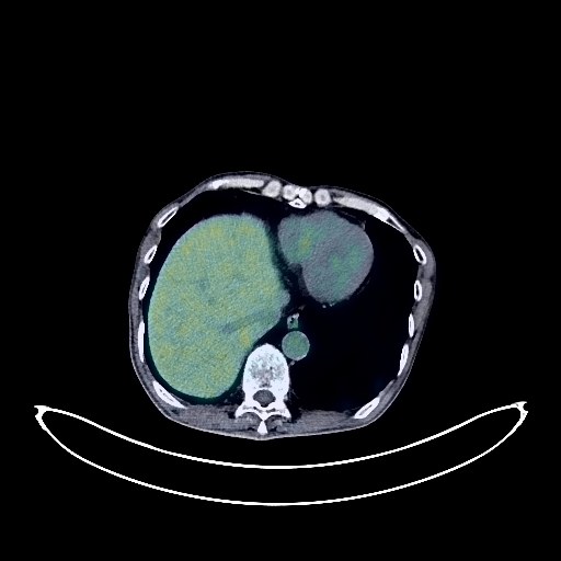
Lung Cancer PET/CT (case 983824-000034 from PETWB-REP)
Whole-body 18F-FDG PET/CT scan in a patient with Lung Cancer taken from the PETWB-REP dataset. The following English report (translated from original Chinese) is taken verbatim from the public dataset and has not been modified or otherwise checked for accuracy (see the end for citation). Impression Right lung cancer after chemotherapy and brain metastasis after radiotherapy: a. Right lower lobe posterior segment lesion with increased FDG metabolism in the solid portion, suggesting tumor activity; multiple lymph node metastases in the right hilum, mediastinum, and right supraclavicular fossa; high probability of right pleural metastasis. b. After brain metastasis radiotherapy, patchy low-density shadow in the right cerebellum with lost FDG uptake; patchy slightly low-density lesions in the right frontal lobe, with relatively uniform density in the temporal lobe, and no abnormalities in FDG metabolism. Considering the changes after treatment, the tumor activity is likely to be basically suppressed; enhanced MRI follow-up is recommended. c. Several small chronic inflammatory nodules (solid) in both lungs. Scattered chronic inflammation and old lesions in both lungs. Emphysema with small bullae in both lungs. Calcification of some arterial walls (including coronary arteries). Localized thickening of the left pleura. Diverticulum in the gastric fundus. Inflammatory or physiological uptake in some intestinal segments. Hemorrhoids. Calcification in the right lobe of the liver. Benign prostatic hyperplasia, with uneven FDG metabolism in the gland; PSA and ultrasound follow-up are recommended. Left scrotal calcification. Degenerative changes in the spine. Multiple Schmorl's nodes in the thoracic and lumbar vertebrae. L4/5 and L5/S1 intervertebral disc bulges. Benign osteopathy of the left iliac bone. This case is from PETWB-REP, a curated dataset of whole-body 18F-FDG PET/CT scans and corresponding radiology reports from 490 patients with a broad spectrum of malignancies. The data were retrospectively collected from patients who underwent clinically indicated whole-body 18F-FDG PET/CT scans at the Shanghai Universal Medical Imaging Diagnostic Center between 2021 and 2024. License: Creative Commons Attribution 4.0 International (CC BY 4.0) Citation: Xue, L., Feng, G., Wenbo, Z., Zhang, Y., Li, L., Wang, S., Peng, L., Peng, S., & Gao, X. (2026). PETWB-REP: A Multi-Cancer Whole-Body FDG PET/CT Dataset with Corresponding Radiology Reports [Data set]. Zenodo. https://doi.org/10.5281/zenodo.18670487
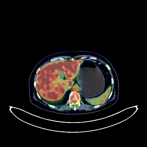
Colon Cancer PET/CT (case 983824-000035 from PETWB-REP)
Whole-body 18F-FDG PET/CT scan in a patient with Colon Cancer taken from the PETWB-REP dataset. The following English report (translated from original Chinese) is taken verbatim from the public dataset and has not been modified or otherwise checked for accuracy (see the end for citation). Impression Post-EMR changes in the colon: No abnormal FDG metabolism was observed in the sigmoid colon metal clip area; no obvious space-occupying lesions were seen in the remaining intestinal segments, and FDG metabolism was normal. Follow-up colonoscopy is recommended. Right upper lobe aberrant lobe formation; chronic inflammatory micronodules in the right upper lobe. A few post-inflammatory lesions in both lungs. Partial arteriosclerosis. Possible hemangioma in the liver; please confirm with contrast-enhanced MRI. Gallbladder bile concentration. Chronic inflammatory changes in the gastric antrum; please confirm with endoscopy. Degenerative changes in the spine; L3/4 intervertebral disc bulge. Widespread increased FDG metabolism in the medullary cavity throughout the body, suggesting possible reactive proliferative changes. No obvious abnormalities were seen on cranial scintigraphy. A few chronic inflammations in the left maxillary sinus. Left upper alveolar ulceritis. This case is from PETWB-REP, a curated dataset of whole-body 18F-FDG PET/CT scans and corresponding radiology reports from 490 patients with a broad spectrum of malignancies. The data were retrospectively collected from patients who underwent clinically indicated whole-body 18F-FDG PET/CT scans at the Shanghai Universal Medical Imaging Diagnostic Center between 2021 and 2024. License: Creative Commons Attribution 4.0 International (CC BY 4.0) Citation: Xue, L., Feng, G., Wenbo, Z., Zhang, Y., Li, L., Wang, S., Peng, L., Peng, S., & Gao, X. (2026). PETWB-REP: A Multi-Cancer Whole-Body FDG PET/CT Dataset with Corresponding Radiology Reports [Data set]. Zenodo. https://doi.org/10.5281/zenodo.18670487
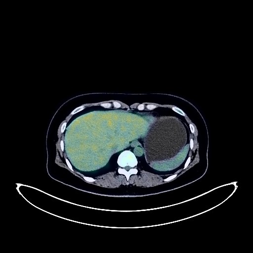
Cervical Cancer PET/CT (case 983824-000036 from PETWB-REP)
Whole-body 18F-FDG PET/CT scan in a patient with Cervical Cancer taken from the PETWB-REP dataset. The following English report (translated from original Chinese) is taken verbatim from the public dataset and has not been modified or otherwise checked for accuracy (see the end for citation). Impression a. Cervical mass with increased FDG metabolism, consistent with cervical cancer; reactive hyperplasia of the left iliac lymph nodes is possible, metastasis to be ruled out, follow-up is recommended. b. Nodular FDG uptake foci on the right pelvic wall, possibly due to physiological uptake by the ureter; lymph nodes to be ruled out, MRI is recommended. Physiological uptake in the uterine cavity. Chronic inflammatory micronodules in the right lung. A few post-inflammatory lesions in both lungs. Anemia. Bilateral breast hyperplasia. Small cyst in the right lobe of the liver. Accessory spleen. Possible hemorrhoids. L3/4, L4/5, and L5/S1 intervertebral disc bulges. Bilateral sacroiliac joint condensation osteitis. No obvious abnormalities were found on cranial scintigraphy. This case is from PETWB-REP, a curated dataset of whole-body 18F-FDG PET/CT scans and corresponding radiology reports from 490 patients with a broad spectrum of malignancies. The data were retrospectively collected from patients who underwent clinically indicated whole-body 18F-FDG PET/CT scans at the Shanghai Universal Medical Imaging Diagnostic Center between 2021 and 2024. License: Creative Commons Attribution 4.0 International (CC BY 4.0) Citation: Xue, L., Feng, G., Wenbo, Z., Zhang, Y., Li, L., Wang, S., Peng, L., Peng, S., & Gao, X. (2026). PETWB-REP: A Multi-Cancer Whole-Body FDG PET/CT Dataset with Corresponding Radiology Reports [Data set]. Zenodo. https://doi.org/10.5281/zenodo.18670487
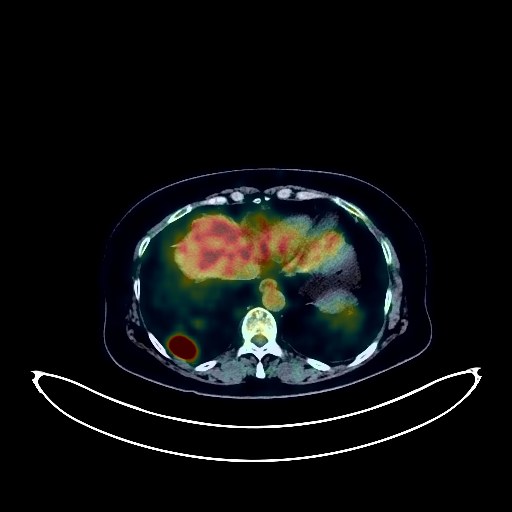
Rectal Cancer PET/CT (case 983824-000037 from PETWB-REP)
Whole-body 18F-FDG PET/CT scan in a patient with Rectal Cancer taken from the PETWB-REP dataset. The following English report (translated from original Chinese) is taken verbatim from the public dataset and has not been modified or otherwise checked for accuracy (see the end for citation). Impression a. Post-rectal cancer surgery, increased FDG metabolism in the lower rectum to anal canal suggests possible inflammatory uptake; tumor recurrence needs to be ruled out. Please have a follow-up colonoscopy. b. Retroperitoneal and right posterior diaphragmatic lymph node metastasis, involving the upper and middle segments of the left ureter, dilation and effusion of the left renal pelvis and upper ureter, and left kidney atrophy. The right kidney shows compensatory slight enlargement. c. Metastasis to the left upper cervical deep space and left supraclavicular lymph nodes. d. Multiple metastatic tumors in both lungs. Multiple bronchiectasis in both lungs, predominantly in the lower lobe of the left lung. Possible mass in the lower part of the left kidney; enhanced MRI is recommended. A few ischemic lesions deep in the brain, senile cerebral changes. Cholestasis in the gallbladder. Fatty infiltration of the pancreas. Osteophyte formation in some vertebrae. This case is from PETWB-REP, a curated dataset of whole-body 18F-FDG PET/CT scans and corresponding radiology reports from 490 patients with a broad spectrum of malignancies. The data were retrospectively collected from patients who underwent clinically indicated whole-body 18F-FDG PET/CT scans at the Shanghai Universal Medical Imaging Diagnostic Center between 2021 and 2024. License: Creative Commons Attribution 4.0 International (CC BY 4.0) Citation: Xue, L., Feng, G., Wenbo, Z., Zhang, Y., Li, L., Wang, S., Peng, L., Peng, S., & Gao, X. (2026). PETWB-REP: A Multi-Cancer Whole-Body FDG PET/CT Dataset with Corresponding Radiology Reports [Data set]. Zenodo. https://doi.org/10.5281/zenodo.18670487
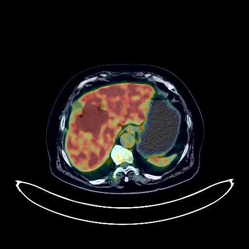
Liver Cancer PET/CT (case 983824-000038 from PETWB-REP)
Whole-body 18F-FDG PET/CT scan in a patient with Liver Cancer taken from the PETWB-REP dataset. The following English report (translated from original Chinese) is taken verbatim from the public dataset and has not been modified or otherwise checked for accuracy (see the end for citation). Impression A mass between the right anterior lobe and left medial lobe of the liver, with increased FDG metabolism, suggestive of malignancy, most likely cholangiocarcinoma. Metastasis to the hilar lymph nodes is also highly probable. Schistosomiasis-related liver disease. Chronic cholecystitis. Gallstones. Post-inferior vena cava filter placement changes. Several small, solid, chronic inflammatory nodules in both lungs. A few chronic inflammations and old lesions in both lungs. A full cardiac silhouette with partial calcification of the arterial walls (including the coronary arteries). Continuous increased FDG metabolism in the sigmoid colon and rectum, suggestive of inflammatory or physiological uptake; colonoscopy follow-up is recommended. Degenerative changes in the spine. L3-S1 intervertebral disc bulge and pneumatosis. Inflammatory or physiological uptake in the right premandibular region; clinical correlation is recommended. A few ischemic lesions in the deep bilateral cerebral regions, suggestive of age-related encephalopathy. Reactive hyperplasia of bilateral cervical lymph nodes. This case is from PETWB-REP, a curated dataset of whole-body 18F-FDG PET/CT scans and corresponding radiology reports from 490 patients with a broad spectrum of malignancies. The data were retrospectively collected from patients who underwent clinically indicated whole-body 18F-FDG PET/CT scans at the Shanghai Universal Medical Imaging Diagnostic Center between 2021 and 2024. License: Creative Commons Attribution 4.0 International (CC BY 4.0) Citation: Xue, L., Feng, G., Wenbo, Z., Zhang, Y., Li, L., Wang, S., Peng, L., Peng, S., & Gao, X. (2026). PETWB-REP: A Multi-Cancer Whole-Body FDG PET/CT Dataset with Corresponding Radiology Reports [Data set]. Zenodo. https://doi.org/10.5281/zenodo.18670487
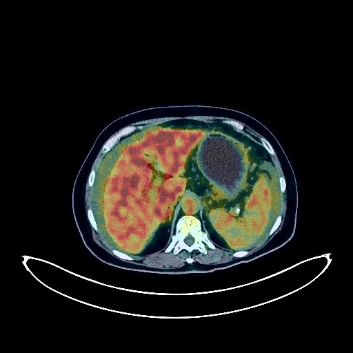
Pancreatic Cancer PET/CT (case 983824-000039 from PETWB-REP)
Whole-body 18F-FDG PET/CT scan in a patient with Pancreatic Cancer taken from the PETWB-REP dataset. The following English report (translated from original Chinese) is taken verbatim from the public dataset and has not been modified or otherwise checked for accuracy (see the end for citation). Impression a. A mass in the neck and body of the pancreas, with elevated FDG metabolism, suggestive of pancreatic cancer. Extensive peritoneal metastasis in the abdominopelvic cavity. b. Metastasis to the peripancreatic, hepatic hilum, retroperitoneum, left supraclavicular fossa, and left posterior cervical triangle lymph nodes. c. Diffuse metastases in both lungs. Abdominal and pelvic effusions. Small amounts of pleural effusion bilaterally. Chronic cholecystitis. Degenerative changes in the spine. L4/5 and L5/S1 intervertebral disc bulges. Post-left total hip replacement surgery changes. Left basal ganglia softening lesion, a few ischemic lesions in the deep bilateral brain regions, senile encephalopathy. This case is from PETWB-REP, a curated dataset of whole-body 18F-FDG PET/CT scans and corresponding radiology reports from 490 patients with a broad spectrum of malignancies. The data were retrospectively collected from patients who underwent clinically indicated whole-body 18F-FDG PET/CT scans at the Shanghai Universal Medical Imaging Diagnostic Center between 2021 and 2024. License: Creative Commons Attribution 4.0 International (CC BY 4.0) Citation: Xue, L., Feng, G., Wenbo, Z., Zhang, Y., Li, L., Wang, S., Peng, L., Peng, S., & Gao, X. (2026). PETWB-REP: A Multi-Cancer Whole-Body FDG PET/CT Dataset with Corresponding Radiology Reports [Data set]. Zenodo. https://doi.org/10.5281/zenodo.18670487
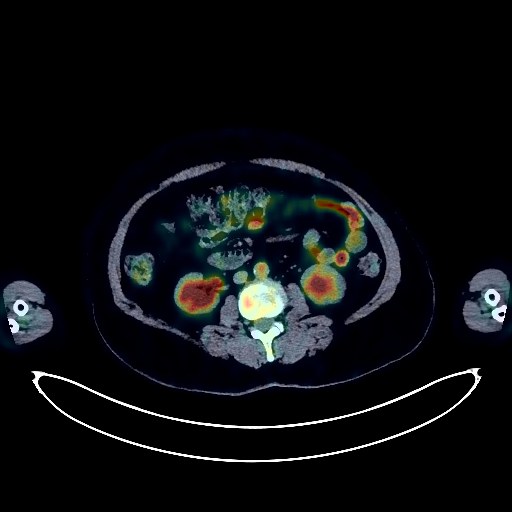
Lymphoma PET/CT (case 983824-000040 from PETWB-REP)
Whole-body 18F-FDG PET/CT scan in a patient with Lymphoma taken from the PETWB-REP dataset. The following English report (translated from original Chinese) is taken verbatim from the public dataset and has not been modified or otherwise checked for accuracy (see the end for citation). Impression a. Space-occupying lesions in the left nasal cavity, left maxillary sinus, and left ethmoid sinus, with increased FDG metabolism, consistent with lymphoma, involving the left orbit, with exophthalmos. b. Hypermetabolic lesions in the spleen, suggestive of lymphoma infiltration. c. Possible infection in the left lower lobe of the lung, lymphoma infiltration to be ruled out; follow-up examination after treatment is recommended for comparison. d. Multiple lymph nodes in both sides of the neck, both hilum, and mediastinum, with increased FDG metabolism, highly suggestive of reactive lymph node hyperplasia; follow-up is recommended to rule out lymphoma infiltration. e. Possible reactive hyperplasia of the entire bone marrow; follow-up is recommended to rule out lymphoma infiltration. A few chronic inflammations and old lesions in both lungs. Calcification of some arterial walls (including coronary arteries). Increased FDG metabolism in the terminal ileum and part of the colon, suggestive of inflammatory or physiological uptake. Duodenal diverticulum in the descending part. Possible uterine cavity effusion, cystic lesions cannot be ruled out; further ultrasound examination is recommended. Degenerative changes in the spine. L4/5 and L5/S1 intervertebral disc bulge. No abnormalities were found on cranial scintigraphy. This case is from PETWB-REP, a curated dataset of whole-body 18F-FDG PET/CT scans and corresponding radiology reports from 490 patients with a broad spectrum of malignancies. The data were retrospectively collected from patients who underwent clinically indicated whole-body 18F-FDG PET/CT scans at the Shanghai Universal Medical Imaging Diagnostic Center between 2021 and 2024. License: Creative Commons Attribution 4.0 International (CC BY 4.0) Citation: Xue, L., Feng, G., Wenbo, Z., Zhang, Y., Li, L., Wang, S., Peng, L., Peng, S., & Gao, X. (2026). PETWB-REP: A Multi-Cancer Whole-Body FDG PET/CT Dataset with Corresponding Radiology Reports [Data set]. Zenodo. https://doi.org/10.5281/zenodo.18670487
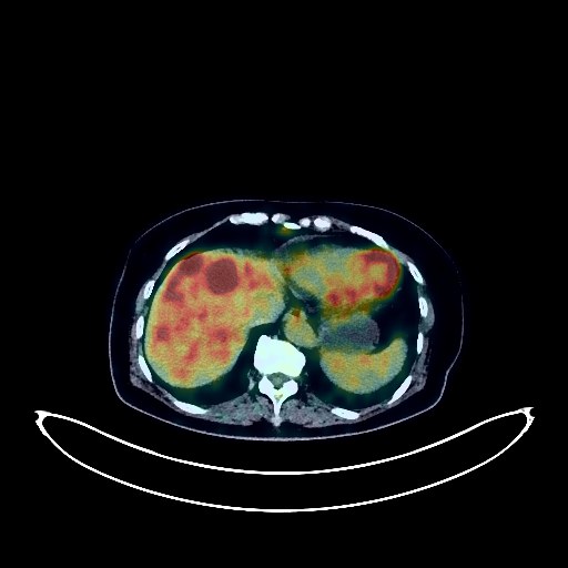
Pancreatic Cancer PET/CT (case 983824-000041 from PETWB-REP)
Whole-body 18F-FDG PET/CT scan in a patient with Pancreatic Cancer taken from the PETWB-REP dataset. The following English report (translated from original Chinese) is taken verbatim from the public dataset and has not been modified or otherwise checked for accuracy (see the end for citation). Impression a. Masses in the neck and body of the pancreas, with increased FDG metabolism, suggestive of pancreatic cancer involving surrounding fat spaces and the celiac trunk. Multiple intrahepatic metastases. Right femoral head metastasis. b. Multiple small lymph nodes between the superior mesenteric artery and the hepatogastric ligament, with mild FDG uptake, suggestive of reactive lymph node hyperplasia; follow-up CT scan recommended to rule out metastasis. c. Strip-like thickening of the pelvic floor peritoneum, with mild FDG uptake; metastasis to be ruled out; clinical correlation recommended. a. Multiple nodules in both lungs, with no abnormal FDG uptake, suggestive of inflammatory nodules, with some mixed metastases to be ruled out; close follow-up CT scan recommended. b. Multiple ground-glass nodules in both lungs, with no abnormal FDG metabolism, suggestive of atypical adenomatous hyperplasia or inflammatory nodules. Small cyst in the left lobe of the liver. Right renal cyst. Left adrenal hyperplasia is likely; follow-up examination recommended. Postoperative absence of the uterus. Spinal degenerative changes, L3/4 and L4/5 intervertebral disc bulging, L4/5 and L5/S1 intervertebral disc pneumatosis and degeneration. This case is from PETWB-REP, a curated dataset of whole-body 18F-FDG PET/CT scans and corresponding radiology reports from 490 patients with a broad spectrum of malignancies. The data were retrospectively collected from patients who underwent clinically indicated whole-body 18F-FDG PET/CT scans at the Shanghai Universal Medical Imaging Diagnostic Center between 2021 and 2024. License: Creative Commons Attribution 4.0 International (CC BY 4.0) Citation: Xue, L., Feng, G., Wenbo, Z., Zhang, Y., Li, L., Wang, S., Peng, L., Peng, S., & Gao, X. (2026). PETWB-REP: A Multi-Cancer Whole-Body FDG PET/CT Dataset with Corresponding Radiology Reports [Data set]. Zenodo. https://doi.org/10.5281/zenodo.18670487
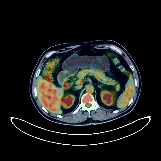
Lung Cancer PET/CT (case 983824-000042 from PETWB-REP)
Whole-body 18F-FDG PET/CT scan in a patient with Lung Cancer taken from the PETWB-REP dataset. The following English report (translated from original Chinese) is taken verbatim from the public dataset and has not been modified or otherwise checked for accuracy (see the end for citation). Impression a. A mass in the lower lobe of the left lung with significantly increased FDG metabolism, suggestive of left lung cancer with surrounding obstructive inflammation. Multiple metastases in both lungs. Metastases to the left hilar, mediastinal, and left supraclavicular fossa lymph nodes. b. Multiple bone metastases in the right ribs, sternum, spine, and pelvis. c. Possible left frontal lobe brain metastasis; enhanced MRI is required for confirmation. Chronic inflammatory changes in the gastric antrum; endoscopic re-examination is necessary if needed. Prostatic calcifications. Left inguinal hernia. Reactive hyperplasia of bilateral inguinal lymph nodes. Degenerative changes in the spine. Reactive hyperplasia of bilateral cervical lymph nodes. This case is from PETWB-REP, a curated dataset of whole-body 18F-FDG PET/CT scans and corresponding radiology reports from 490 patients with a broad spectrum of malignancies. The data were retrospectively collected from patients who underwent clinically indicated whole-body 18F-FDG PET/CT scans at the Shanghai Universal Medical Imaging Diagnostic Center between 2021 and 2024. License: Creative Commons Attribution 4.0 International (CC BY 4.0) Citation: Xue, L., Feng, G., Wenbo, Z., Zhang, Y., Li, L., Wang, S., Peng, L., Peng, S., & Gao, X. (2026). PETWB-REP: A Multi-Cancer Whole-Body FDG PET/CT Dataset with Corresponding Radiology Reports [Data set]. Zenodo. https://doi.org/10.5281/zenodo.18670487
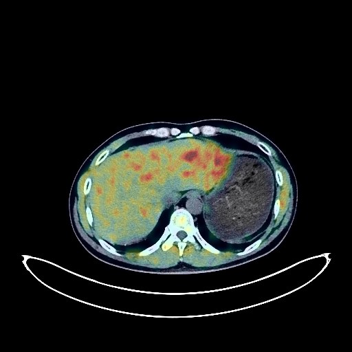
Lung Cancer PET/CT (case 983824-000043 from PETWB-REP)
Whole-body 18F-FDG PET/CT scan in a patient with Lung Cancer taken from the PETWB-REP dataset. The following English report (translated from original Chinese) is taken verbatim from the public dataset and has not been modified or otherwise checked for accuracy (see the end for citation). Impression Postoperative changes following a left brain lesion; a slightly high-density nodule in the left temporoparietal-occipital lobe with increased FDG metabolism, suggestive of a metastatic tumor. a. Lung cancer in the apical-posterior segment of the left upper lobe; lung cancer or metastatic lymph nodes in the left hilar region. b. Chronic inflammation and sequelae in both lungs. Paraseptal emphysema in both lungs. Mild osteophyte formation in some vertebral bodies. L4/5 intervertebral disc bulge. This case is from PETWB-REP, a curated dataset of whole-body 18F-FDG PET/CT scans and corresponding radiology reports from 490 patients with a broad spectrum of malignancies. The data were retrospectively collected from patients who underwent clinically indicated whole-body 18F-FDG PET/CT scans at the Shanghai Universal Medical Imaging Diagnostic Center between 2021 and 2024. License: Creative Commons Attribution 4.0 International (CC BY 4.0) Citation: Xue, L., Feng, G., Wenbo, Z., Zhang, Y., Li, L., Wang, S., Peng, L., Peng, S., & Gao, X. (2026). PETWB-REP: A Multi-Cancer Whole-Body FDG PET/CT Dataset with Corresponding Radiology Reports [Data set]. Zenodo. https://doi.org/10.5281/zenodo.18670487
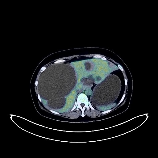
Cervical Cancer PET/CT (case 983824-000044 from PETWB-REP)
Whole-body 18F-FDG PET/CT scan in a patient with Cervical Cancer taken from the PETWB-REP dataset. The following English report (translated from original Chinese) is taken verbatim from the public dataset and has not been modified or otherwise checked for accuracy (see the end for citation). Impression Cervical mass with increased FDG metabolism, involving adjacent uterine body and vagina, consistent with cervical cancer. Multiple lymph node metastases in bilateral iliac vessels, bilateral pelvic walls, para-aortic region, posterior diaphragmatic crura, left infradeep cervical space, left posterior cervical triangle, and left supraclavicular fossa. Ground-glass nodule in the anterior segment of the right upper lobe, FDG metabolism normal, suggestive of inflammatory nodule or atypical adenomatous hyperplasia; annual HRCT follow-up recommended. Calcification in the right middle lobe. Bilateral breast proliferative changes; several soft tissue nodules in both breasts, FDG metabolism normal, suggestive of fibroadenoma or proliferative nodules; ultrasound follow-up recommended. Multiple liver cysts. Right kidney stone or calcification. Complex cyst in the left kidney. Increased FDG metabolism in parts of the intestine, suggestive of inflammatory or physiological uptake. Degenerative changes in the spine. L4/5 and L5/S1 intervertebral disc bulges. A subcutaneous soft tissue nodule in the midline of the buttock; FDG metabolism was normal, suggestive of a sebaceous cyst. Cranial scintigraphy showed no abnormalities. A small amount of chronic inflammation was observed in the left maxillary sinus. This case is from PETWB-REP, a curated dataset of whole-body 18F-FDG PET/CT scans and corresponding radiology reports from 490 patients with a broad spectrum of malignancies. The data were retrospectively collected from patients who underwent clinically indicated whole-body 18F-FDG PET/CT scans at the Shanghai Universal Medical Imaging Diagnostic Center between 2021 and 2024. License: Creative Commons Attribution 4.0 International (CC BY 4.0) Citation: Xue, L., Feng, G., Wenbo, Z., Zhang, Y., Li, L., Wang, S., Peng, L., Peng, S., & Gao, X. (2026). PETWB-REP: A Multi-Cancer Whole-Body FDG PET/CT Dataset with Corresponding Radiology Reports [Data set]. Zenodo. https://doi.org/10.5281/zenodo.18670487
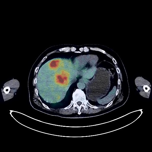
Liver Cancer PET/CT (case 983824-000045 from PETWB-REP)
Whole-body 18F-FDG PET/CT scan in a patient with Liver Cancer taken from the PETWB-REP dataset. The following English report (translated from original Chinese) is taken verbatim from the public dataset and has not been modified or otherwise checked for accuracy (see the end for citation). Impression a. Multiple space-occupying lesions in the liver with increased FDG metabolism, suggestive of malignancy, possibly hepatocellular carcinoma with intrahepatic metastasis, metastatic tumors cannot be ruled out; multiple lymph node metastases in the hilar region and retroperitoneum. b. Multiple bone metastases throughout the body. Pathological fracture of the L4 vertebral body. c. Gallstones with chronic cholecystitis. Increased local FDG uptake in the gallbladder, gallbladder cancer to be ruled out, please combine with enhanced MRI for comprehensive analysis. Benign prostatic hyperplasia with calcification, uneven FDG metabolism, please follow up with PSA and MRI. Chronic inflammatory nodules in the upper lobe of the right lung. Scattered post-inflammatory lesions in both lungs. Anemia changes, calcification of some arterial walls (including coronary arteries). Cyst in the right lobe of the liver. Adrenal hyperplasia on the left side. Chronic inflammatory changes in the antrum of the stomach. Osteoporosis, degenerative changes in the spine, multiple intervertebral disc bulges with pneumoconiosis. Postoperative changes in the left femur. The thyroid gland has uneven density and multiple nodules in both lobes. FDG metabolism is normal, suggesting nodular goiter. Please confirm with ultrasound examination. Elderly patient with deep lacunar infarcts. Please confirm with MRI examination. Right maxillary sinus submucosal cyst. This case is from PETWB-REP, a curated dataset of whole-body 18F-FDG PET/CT scans and corresponding radiology reports from 490 patients with a broad spectrum of malignancies. The data were retrospectively collected from patients who underwent clinically indicated whole-body 18F-FDG PET/CT scans at the Shanghai Universal Medical Imaging Diagnostic Center between 2021 and 2024. License: Creative Commons Attribution 4.0 International (CC BY 4.0) Citation: Xue, L., Feng, G., Wenbo, Z., Zhang, Y., Li, L., Wang, S., Peng, L., Peng, S., & Gao, X. (2026). PETWB-REP: A Multi-Cancer Whole-Body FDG PET/CT Dataset with Corresponding Radiology Reports [Data set]. Zenodo. https://doi.org/10.5281/zenodo.18670487
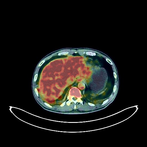
Lung Cancer PET/CT (case 983824-000046 from PETWB-REP)
Whole-body 18F-FDG PET/CT scan in a patient with Lung Cancer taken from the PETWB-REP dataset. The following English report (translated from original Chinese) is taken verbatim from the public dataset and has not been modified or otherwise checked for accuracy (see the end for citation). Impression a. A mass in the right upper lobe with increased FDG uptake; multiple enlarged lymph nodes in the right hilum, mediastinum, and bilateral supraclavicular fossa, some fused, with increased FDG metabolism, suggesting a high probability of right lung cancer with multiple lymph node metastases; tuberculosis to be ruled out; airway dissemination in the right upper lobe is also a possibility. b. Small lymph nodes in the hepatogastric interspace with increased FDG metabolism, suggesting reactive hyperplasia. Small amount of pleural effusion and pericardial effusion bilaterally. a. Ground-glass nodule in the left upper lobe, with normal FDG metabolism, suggesting atypical adenomatous hyperplasia or inflammatory nodule; annual HRCT follow-up is recommended. b. Multiple small chronic inflammatory nodules (solid) in the remaining lungs. Paraseptal emphysema in both upper lobes. Minor inflammation in the bilateral ethmoid and maxillary sinuses. Increased FDG uptake in the left maxillary alveolar region, suggesting inflammation. No obvious abnormalities were seen on cranial imaging. Multiple cysts in both kidneys. Benign prostatic hyperplasia with calcifications. Bilateral testicular hydrocele with small amount of fluid in the tunica vaginalis. Spinal osteophyte formation, L4/5 and L5/S1 intervertebral disc bulge. This case is from PETWB-REP, a curated dataset of whole-body 18F-FDG PET/CT scans and corresponding radiology reports from 490 patients with a broad spectrum of malignancies. The data were retrospectively collected from patients who underwent clinically indicated whole-body 18F-FDG PET/CT scans at the Shanghai Universal Medical Imaging Diagnostic Center between 2021 and 2024. License: Creative Commons Attribution 4.0 International (CC BY 4.0) Citation: Xue, L., Feng, G., Wenbo, Z., Zhang, Y., Li, L., Wang, S., Peng, L., Peng, S., & Gao, X. (2026). PETWB-REP: A Multi-Cancer Whole-Body FDG PET/CT Dataset with Corresponding Radiology Reports [Data set]. Zenodo. https://doi.org/10.5281/zenodo.18670487
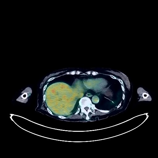
Cholangiocarcinoma PET/CT (case 983824-000047 from PETWB-REP)
Whole-body 18F-FDG PET/CT scan in a patient with Cholangiocarcinoma taken from the PETWB-REP dataset. The following English report (translated from original Chinese) is taken verbatim from the public dataset and has not been modified or otherwise checked for accuracy (see the end for citation). Impression a. Post-PTCD, increased FDG uptake along the indwelling catheter suggests possible post-operative changes, but local tumor uptake cannot be ruled out; no clear space-occupying lesion was seen in the common bile duct area, but intrahepatic bile duct dilation was observed. The above findings should be analyzed in conjunction with enhanced MRI images. b. A metastatic tumor in the left pubic tubercle is highly probable; follow-up is recommended. Reactive hyperplasia of small lymph nodes in the retroperitoneum and bilateral iliac vessels is observed. c. Gallstones or calcifications in the gallbladder, chronic cholecystitis, without clear space-occupying lesions. Uneven density between the left and right lobes of the thyroid gland; slight enlargement of the right thyroid lobe with slightly low-density nodules, increased FDG metabolism, suggesting adenoma; malignancy needs to be ruled out. Further ultrasound examination is recommended, with biopsy if necessary. a. Ground-glass opacity in the apical segment of the right upper lobe, with normal FDG metabolism, suggests chronic inflammatory changes or atypical adenomatous hyperplasia. A follow-up HRCT scan every six months is recommended. b. Chronic inflammatory nodules and nodular lesions in both lungs. A few chronic inflammatory lesions and sequelae in both lungs. Slight thickening of the pleura on both sides, with a small amount of pleural effusion in both pleural cavities. Calcification of some arterial walls (including coronary arteries). Cystic lesions in the neck of the pancreas. Left adrenal hyperplasia. Calcification of the uterus. Slight thickening of the gastric antrum wall with increased FDG metabolism, and increased FDG metabolism in some intestinal segments, suggestive of chronic inflammatory changes or physiological uptake. Osteoporosis, scoliosis with degenerative changes, Schmorl's nodes in the T12 and L2 vertebral bodies. Intervertebral disc bulges at L3/4, L4/5, and L5/S1. Left hip periarthritis. Left sacral wing fracture due to failure. Bilateral deep cerebral ischemic lesions, age-related brain changes. This case is from PETWB-REP, a curated dataset of whole-body 18F-FDG PET/CT scans and corresponding radiology reports from 490 patients with a broad spectrum of malignancies. The data were retrospectively collected from patients who underwent clinically indicated whole-body 18F-FDG PET/CT scans at the Shanghai Universal Medical Imaging Diagnostic Center between 2021 and 2024. License: Creative Commons Attribution 4.0 International (CC BY 4.0) Citation: Xue, L., Feng, G., Wenbo, Z., Zhang, Y., Li, L., Wang, S., Peng, L., Peng, S., & Gao, X. (2026). PETWB-REP: A Multi-Cancer Whole-Body FDG PET/CT Dataset with Corresponding Radiology Reports [Data set]. Zenodo. https://doi.org/10.5281/zenodo.18670487
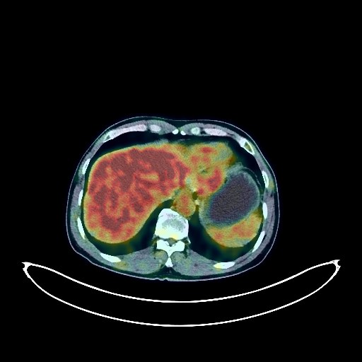
Lung Cancer PET/CT (case 983824-000048 from PETWB-REP)
Whole-body 18F-FDG PET/CT scan in a patient with Lung Cancer taken from the PETWB-REP dataset. The following English report (translated from original Chinese) is taken verbatim from the public dataset and has not been modified or otherwise checked for accuracy (see the end for citation). Impression a. A mass near the hilum in the right upper lobe of the lung, with increased FDG metabolism, suggestive of central lung cancer with obstructive inflammation or airway dissemination. Right hilar lymph node metastasis. Reactive hyperplasia of the left hilar and mediastinal lymph nodes is highly probable; follow-up is recommended. b. Scattered inflammation in the remaining lungs. Calcification of some arterial walls (including coronary arteries). Left renal cyst. Slight thickening of the walls of part of the gastric body and antrum, with mildly increased FDG uptake, suggestive of chronic gastritis; follow-up with gastroscopy is recommended. Degenerative changes in the spine. L4/5 and L5/S1 intervertebral disc bulge. Right shoulder periarthritis. Multiple nodules in both parotid glands, with increased FDG metabolism, suggestive of adenolymphoma; follow-up with MRI is recommended. A few ischemic lesions in the deep bilateral brain regions, suggestive of senile encephalopathy. Chronic inflammation of the left maxillary sinus. This case is from PETWB-REP, a curated dataset of whole-body 18F-FDG PET/CT scans and corresponding radiology reports from 490 patients with a broad spectrum of malignancies. The data were retrospectively collected from patients who underwent clinically indicated whole-body 18F-FDG PET/CT scans at the Shanghai Universal Medical Imaging Diagnostic Center between 2021 and 2024. License: Creative Commons Attribution 4.0 International (CC BY 4.0) Citation: Xue, L., Feng, G., Wenbo, Z., Zhang, Y., Li, L., Wang, S., Peng, L., Peng, S., & Gao, X. (2026). PETWB-REP: A Multi-Cancer Whole-Body FDG PET/CT Dataset with Corresponding Radiology Reports [Data set]. Zenodo. https://doi.org/10.5281/zenodo.18670487
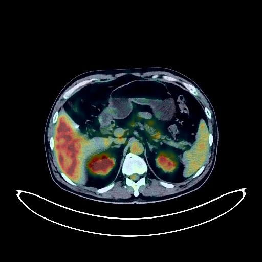
Lung Cancer PET/CT (case 983824-000049 from PETWB-REP)
Whole-body 18F-FDG PET/CT scan in a patient with Lung Cancer taken from the PETWB-REP dataset. The following English report (translated from original Chinese) is taken verbatim from the public dataset and has not been modified or otherwise checked for accuracy (see the end for citation). Impression a. Mass in the lower lobe of the left lung, with increased FDG metabolism, consistent with lung cancer. b. Multiple lymph nodes in the right hilum and mediastinum with some showing increased FDG metabolism, suggesting possible reactive lymph node hyperplasia; partial metastasis is pending. Please compare with previous scans for follow-up. c. Interstitial lung inflammation in both lungs. Bullae in the lower lobes of both lungs. Pleural thickening bilaterally. Multiple calcifications in the right hilum. Calcification of some arterial walls. Small kidney stone in the right kidney. Degenerative changes in the spine. No obvious abnormalities seen on cranial scintigraphy. Bilateral maxillary sinusitis. This case is from PETWB-REP, a curated dataset of whole-body 18F-FDG PET/CT scans and corresponding radiology reports from 490 patients with a broad spectrum of malignancies. The data were retrospectively collected from patients who underwent clinically indicated whole-body 18F-FDG PET/CT scans at the Shanghai Universal Medical Imaging Diagnostic Center between 2021 and 2024. License: Creative Commons Attribution 4.0 International (CC BY 4.0) Citation: Xue, L., Feng, G., Wenbo, Z., Zhang, Y., Li, L., Wang, S., Peng, L., Peng, S., & Gao, X. (2026). PETWB-REP: A Multi-Cancer Whole-Body FDG PET/CT Dataset with Corresponding Radiology Reports [Data set]. Zenodo. https://doi.org/10.5281/zenodo.18670487